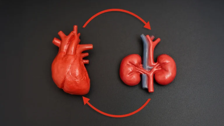


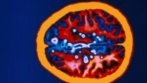


In the silent, intricate architecture of the human brain, a sophisticated plumbing system works tirelessly to sweep away metabolic debris. When this system fails, the consequences are profound. Scientists from Nanyang Technological University, Singapore (NTU Singapore) have unveiled a breakthrough discovery: the brain’s waste-removal pathways often become obstructed in individuals exhibiting the earliest signs of Alzheimer’s disease. These blockages, known as "enlarged perivascular spaces," appear to serve as a critical early-warning biological marker, signaling the onset of cognitive decline long before the devastating symptoms of dementia manifest.
This finding, published following extensive research, offers a potential paradigm shift in how clinicians approach the diagnosis of the world’s most common form of dementia. By utilizing routine magnetic resonance imaging (MRI) scans, doctors may soon be able to identify these "clogged drains" as a standard part of diagnostic protocols, providing a window of opportunity for early intervention that was previously inaccessible.
The Mechanics of Brain Clearance
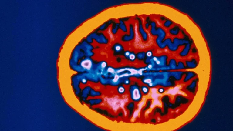
To understand the significance of this discovery, one must first understand the anatomy of the brain’s cleansing mechanism. Encircling the brain’s blood vessels are microscopic channels known as perivascular spaces. These pathways act as a critical drainage system, responsible for flushing out toxic metabolic byproducts—specifically beta-amyloid and tau proteins.
In a healthy brain, these proteins are cleared efficiently. However, in patients developing Alzheimer’s, these waste products accumulate, forming plaques and tangles that disrupt cellular communication. When the drainage system becomes inefficient, these perivascular spaces widen, becoming visibly enlarged on high-resolution MRI scans. While clinicians have long observed these spaces, their direct correlation to the pathology of Alzheimer’s had remained a subject of intense scientific debate until now.
A Study of Global Significance: The Asian Perspective
One of the most compelling aspects of the NTU Singapore study is its deliberate focus on Asian populations. For decades, Alzheimer’s research has been heavily skewed toward Caucasian cohorts, creating a "data gap" that limits the universal applicability of diagnostic criteria.
Associate Professor Nagaendran Kandiah, who led the study at NTU’s Lee Kong Chian School of Medicine (LKCMedicine), notes that the genetic and physiological landscape of dementia can vary significantly across ethnicities. For instance, the apolipoprotein E4 (APOE4) gene—a major risk factor for Alzheimer’s—is present in approximately 50 to 60 percent of Caucasian dementia patients. In contrast, among Singaporean patients, that figure drops below 20 percent.
By analyzing a cohort of nearly 1,000 individuals in Singapore—a population reflecting a rich tapestry of ethnic backgrounds—the NTU team ensured that their findings were grounded in a demographic reality that is often ignored. This inclusive approach underscores the necessity of region-specific medical research, ensuring that diagnostic tools developed in one part of the world are validated and effective for populations globally.
Chronology of the Investigation
The research was conducted as part of the Scholarly Project module within LKCMedicine’s Bachelor of Medicine and Bachelor of Surgery programme. The methodology was structured to establish a clear causal link between structural brain changes and biochemical markers of disease:
- Participant Selection: The study cohort comprised nearly 350 individuals with normal cognitive function and a larger group exhibiting signs of mild cognitive impairment (MCI). MCI is recognized as a "pre-dementia" state, where individuals experience noticeable memory or thinking difficulties that do not yet meet the threshold for a full dementia diagnosis.
- Structural Mapping: Researchers utilized MRI scans to quantify the presence and size of perivascular spaces, mapping these against known indicators of brain health, such as white matter integrity—the nerve fiber network that facilitates inter-regional communication.
- Biochemical Correlation: The team cross-referenced these scans with blood-based biomarkers. By measuring seven distinct Alzheimer’s-related biochemicals, including levels of beta-amyloid and tau proteins, the scientists were able to create a multidimensional view of the participants’ brain health.
- Comparative Analysis: The data revealed that participants with MCI were significantly more likely to display enlarged perivascular spaces than their cognitively healthy counterparts. Furthermore, the correlation between these clogged pathways and Alzheimer’s-related biochemical markers proved to be stronger than the correlation with white matter damage—a traditional diagnostic marker.
Supporting Data: Why "Clogged Drains" Matter More
The study’s most striking finding lies in the comparative strength of its indicators. While white matter damage has long been the "gold standard" for evaluating cerebrovascular contributions to dementia, the NTU research suggests it may be a lagging indicator.
When researchers mapped the seven blood markers against structural brain changes, they found that enlarged perivascular spaces were associated with four of the seven indicators. Most importantly, among the MCI group, the association between these biochemical markers and enlarged perivascular spaces was more potent than the association with white matter damage. This suggests that the obstruction of the brain’s drainage system is a primary, early-stage event in the cascade of Alzheimer’s pathology.
Expert Perspectives and Official Responses
The implications of this study have been met with cautious optimism by the medical community. Dr. Rachel Cheong Chin Yee, a Senior Consultant at Khoo Teck Puat Hospital, emphasizes the clinical utility of these findings. "These findings are significant because they suggest that brain scans showing enlarged perivascular spaces could potentially help identify people at higher risk of Alzheimer’s disease, even before symptoms appear," Dr. Cheong noted.
Dr. Chong Yao Feng, a Consultant at the National University Hospital, adds that the study challenges the traditional separation of cerebrovascular disease and Alzheimer’s. "The study’s findings are intriguing as they demonstrate that both diseases do interact in a synergistic manner," Dr. Chong explained. He cautions, however, that clinical judgment remains paramount. Doctors must synthesize MRI data with patient history and symptoms, as the presence of enlarged spaces does not inherently guarantee an Alzheimer’s diagnosis, but rather signals a need for further, targeted investigation.
Justin Ong, the study’s lead author and a fifth-year medical student, highlights the ethical and practical necessity of this research. By identifying patients at risk earlier, doctors gain a critical window to intervene—whether through lifestyle modifications, pharmacological trials, or closer monitoring—potentially slowing the progression of memory loss and mood changes that strip patients of their independence.
Clinical Implications: A Path to Early Intervention
The "clinical implications" cited by Associate Professor Kandiah are vast. Currently, early detection of Alzheimer’s is often hindered by the high cost and invasiveness of advanced diagnostic tests, such as cerebrospinal fluid analysis or specialized PET scans. If routine MRI scans—which are already performed for patients reporting cognitive decline—can be leveraged to detect these specific structural anomalies, the barrier to early diagnosis could be significantly lowered.
Furthermore, this discovery provides a new target for therapeutic intervention. If the "clogging" of these spaces is indeed a driver of disease, future research may explore whether improving the efficiency of the glymphatic system (the brain’s waste clearance system) could be a viable strategy to slow or prevent the development of Alzheimer’s.
Future Trajectories: What Lies Ahead
The NTU research team is not resting on these initial findings. The next phase of the project involves longitudinal tracking of the participants. By observing how many of those with enlarged perivascular spaces progress to dementia over time, the researchers aim to solidify the predictive power of this marker.
If these results are validated in broader international studies, the routine identification of enlarged perivascular spaces could become a standard component of neurologists’ diagnostic checklists. This would represent a major victory in the fight against dementia, moving the medical community away from a reactive model—where treatment begins only after symptoms are debilitating—toward a proactive, preventative model.
As the population ages, the global burden of Alzheimer’s continues to rise. Breakthroughs that leverage existing technologies, such as the humble MRI, to uncover the hidden mechanisms of the disease are exactly what is needed to change the narrative. The work of the NTU Singapore team serves as a beacon of progress, illuminating the microscopic pathways of the brain and, in doing so, offering hope to millions of families worldwide.