









Featured Buzz – January 11, 2026
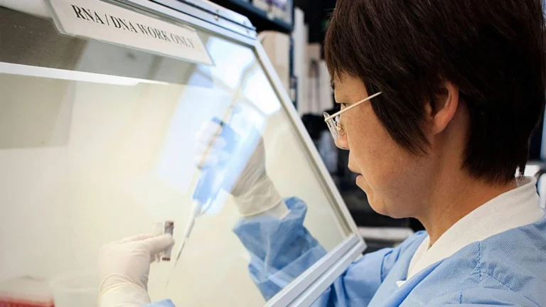
The landscape of respiratory medicine is undergoing a seismic shift as clinicians increasingly turn to artificial intelligence (AI) and advanced imaging to solve some of the most persistent diagnostic challenges. From the routine electrocardiogram (ECG) to the high-stakes environment of the Intensive Care Unit (ICU), three new studies—published in eBioMedicine, the Journal of Perinatology, and Nature Communications—illustrate how technology is bridging the gap between clinical suspicion and definitive diagnosis.
By repurposing standard diagnostic tools and integrating cutting-edge biomarkers, researchers are providing clinicians with the precision necessary to intervene earlier, reduce mortality, and curb the global crisis of antibiotic overuse.
I. ECGs and AI: A New Frontier for COPD Screening
Chronic Obstructive Pulmonary Disease (COPD) remains one of the world’s leading causes of morbidity and mortality, yet it is notoriously underdiagnosed. Millions live with compromised lung function, often only seeking medical attention once the disease has progressed to a debilitating stage.
The Mechanism: Repurposing the ECG
Researchers from the Mount Sinai Health System have introduced a paradigm-shifting approach: using AI-driven Convolutional Neural Networks (CNNs) to identify COPD patterns within standard electrocardiograms. While ECGs are traditionally used to monitor cardiac electrical activity, the researchers hypothesized that the subtle physiological shifts caused by chronic lung disease—such as pulmonary hypertension and right ventricular strain—leave a digital "fingerprint" on the heart’s electrical tracing.
Chronology and Methodology
The study was massive in scale, reflecting a modern approach to big data in medicine. The investigators compiled a dataset of 208,231 ECGs from 18,225 patients with confirmed COPD across five Mount Sinai hospitals in the New York metropolitan area. To ensure accuracy, they compared this against a control group of 552,771 ECGs from 59,356 individuals, meticulously matched by age, sex, and race.
The model was subjected to rigorous validation, moving from internal testing to two distinct external validation cohorts. This included a final test group of 258 COPD patients and 1,290 matched controls, ensuring the algorithm’s performance was not merely a result of overfitting to specific institutional data.
Implications for Primary Care
While the researchers are quick to emphasize that AI-enhanced ECGs will not replace gold-standard spirometry, the implications are profound. Integrating this model into routine clinical visits could serve as a "red flag" system. If a patient’s ECG suggests a high probability of COPD, clinicians can initiate timely smoking cessation, introduce pulmonary rehabilitation, and optimize targeted therapies. This early intervention could drastically slow disease progression and reduce the long-term healthcare burden on both patients and the hospital system.
II. Predicting Extubation Success in Preterm Infants
In the Neonatal Intensive Care Unit (NICU), the decision to extubate a very low birth weight (VLBW) infant is a critical juncture. Premature extubation can lead to respiratory distress and the trauma of reintubation, while delayed extubation increases the risk of ventilator-associated pneumonia and prolonged hospitalization.
The Role of Lung Ultrasound (LUS)
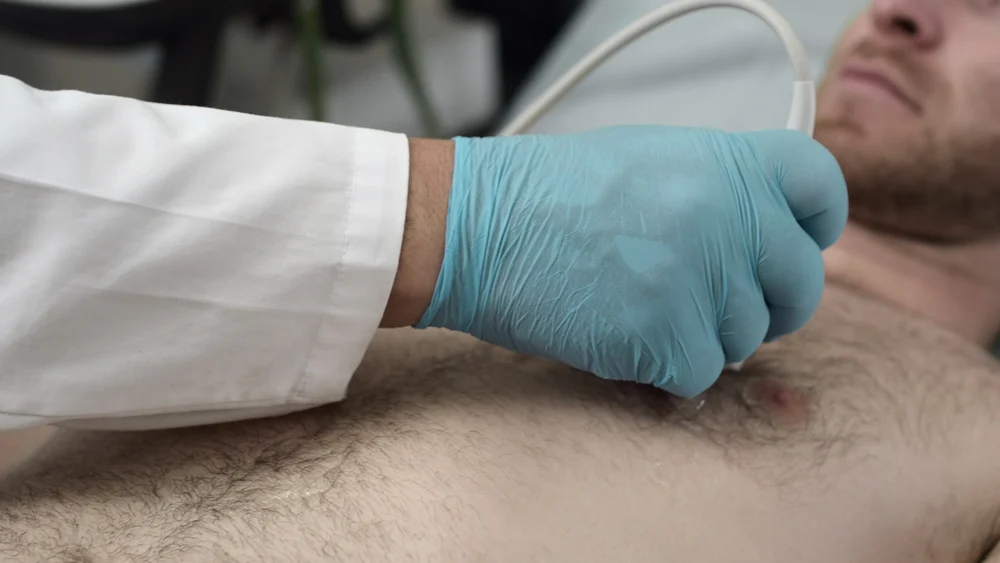
A team from the University of Chicago has demonstrated that lung ultrasound (LUS) scores offer a predictive accuracy previously unattainable with standard clinical assessment alone. By monitoring the aerated state of the lungs in real-time, clinicians can better gauge whether an infant is physiologically ready to breathe independently.
Supporting Data
The study tracked 45 VLBW infants who had been intubated due to respiratory distress syndrome, covering 53 distinct extubation attempts. The protocol required a LUS to be performed three to six hours prior to each attempt. A "failed" extubation was strictly defined as any instance requiring reintubation within seven days.
The results underscored the efficacy of the neonatal-adapted LUS score. Notably, the study also provided clarity on pharmacological support, finding that dexamethasone treatment—often used to reduce inflammation—did not negatively correlate with LUS scores, providing clinicians with confidence in using the score regardless of recent steroid interventions.
Official Perspectives
The researchers assert that the LUS score is now a vital component of the neonatal "toolbox." By performing these scans on the day of extubation, pediatricians can move away from "trial-by-fire" extubation methods and toward a data-driven approach that minimizes the physical stress placed on the most vulnerable patients.
III. AI, Biomarkers, and the War on Antibiotic Misuse
Perhaps the most ambitious study comes from the University of California, San Francisco (UCSF), where researchers are tackling the over-prescription of antibiotics in critically ill patients with Lower Respiratory Tract Infections (LRTIs).
The Diagnostic Strategy: FABP4 and Generative AI
The UCSF team developed a dual-layered diagnostic strategy. First, they focused on a specific biomarker, FABP4 (Fatty Acid Binding Protein 4), found in lung fluid. FABP4 acts as an inflammatory regulator; its expression is significantly diminished in infected lung cells compared to healthy ones.
Second, they fed this biological data into a generative AI model, which also processed information from electronic medical records (EMRs), including chest X-ray reports and clinical notes from the bedside medical team.
Chronology and Results
In an observational study involving critically ill adults, the model achieved a 96% diagnostic accuracy rate, consistently outperforming the subjective assessments of ICU clinicians. The findings were stark: in the study cohort, many clinicians were inclined to prescribe broad-spectrum antibiotics "just in case." The researchers believe that by relying on the AI-FABP4 integration, the use of inappropriate antibiotics could have been reduced by over 80%.
Future Directions
The UCSF team is currently moving toward validating the model as a formal clinical test. Their vision extends beyond pneumonia; they plan to adapt the architecture of this AI to assist in the early diagnosis of sepsis, a condition where every hour of delayed or inappropriate treatment carries a significant mortality risk.
IV. Synthesis and Future Outlook
The common thread linking these three studies is the move away from clinical intuition toward data-augmented precision. Whether it is a CNN identifying the subtle heart-lung connection in an ECG, a neonatologist using ultrasound to visualize lung compliance, or an ICU physician using a generative AI to interpret complex biomarkers, the goal remains the same: Precision Medicine.
Professional and Ethical Implications
The integration of AI into these workflows raises important questions regarding the role of the human clinician. However, the researchers in all three studies are aligned: AI is not a replacement for the physician, but an advanced tool for synthesis. By distilling massive amounts of data—ECG waveforms, LUS imagery, and multi-modal medical records—these tools allow doctors to focus on the human elements of care: the bedside consultation, the family discussion, and the long-term management of complex diseases.
The Road Ahead
As these technologies transition from research papers to bedside applications, the healthcare industry faces the challenge of integration. Hospitals must ensure that these models are HIPAA-compliant, transparent, and accessible within existing EMR infrastructures.
Furthermore, the success of these studies reinforces the necessity of interdisciplinary collaboration. The marriage of engineering, data science, and clinical respiratory medicine has yielded results that were considered futuristic just a decade ago. If the trajectory of these trials continues, we are entering an era where diagnostic errors in respiratory health may become the exception rather than the norm.
Conclusion
From the NICU to the adult ICU, the ability to predict, identify, and treat respiratory disease is being fundamentally rewritten. As we move through 2026, the focus will likely shift from "can we do this?" to "how quickly can we scale this?" For patients, this means the promise of shorter hospital stays, fewer complications, and a significantly higher quality of life. The data is in, the models are validated, and the future of respiratory diagnostics is clearly powered by the synergy between human expertise and machine intelligence.