











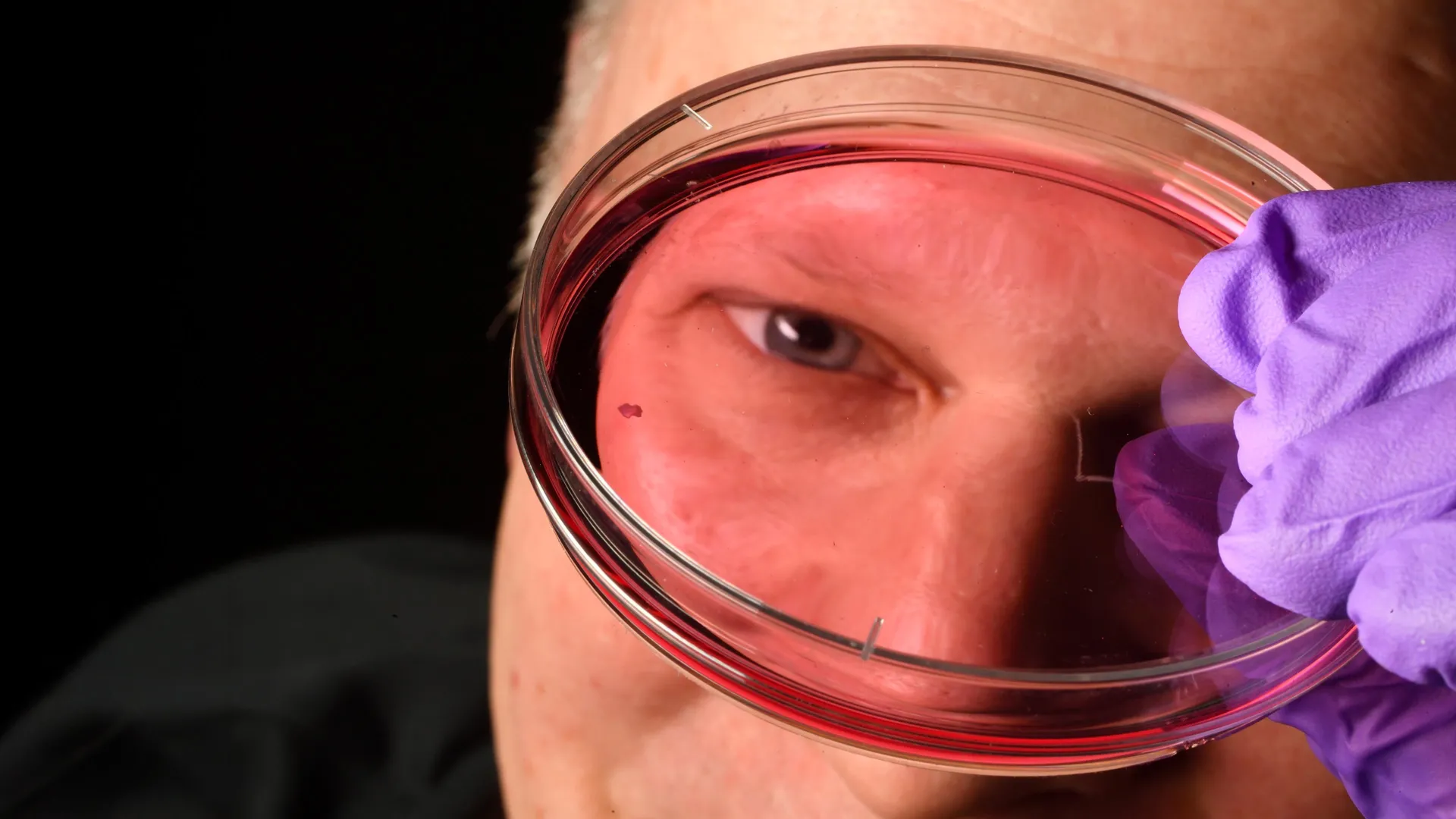
In a breakthrough that fundamentally alters our understanding of human ocular development, researchers at Johns Hopkins University have identified the biological "switch" that enables the formation of the foveola—the tiny, specialized center of the retina responsible for our sharpest, most detailed vision. By utilizing cutting-edge retinal organoid technology, the team has successfully mapped the precise molecular choreography that occurs in the womb, effectively overturning a three-decade-old scientific consensus regarding how photoreceptor cells achieve their unique arrangement.
This discovery, published in the Proceedings of the National Academy of Sciences, not only solves a long-standing mystery in developmental biology but also paves a high-stakes pathway toward regenerative medicine. By decoding the mechanism by which the eye fine-tunes its color-sensing capabilities, scientists are now one step closer to developing cell-replacement therapies for debilitating conditions such as macular degeneration and glaucoma, for which there are currently few, if any, curative options.
The Foveola: The High-Definition Center of Perception
To understand the magnitude of this discovery, one must appreciate the anatomy of the human eye. The foveola is a sub-region within the fovea, the central pit of the retina. Although this region occupies a minute fraction of the total retinal surface area, it is the powerhouse of human visual perception, accounting for approximately 50% of our visual processing.
The retina is carpeted with photoreceptor cells known as cones, which are responsible for daytime and color vision. Humans are trichromatic, meaning we possess three distinct types of cones sensitive to blue, green, and red wavelengths of light. However, the distribution of these cells is not uniform. While the periphery of the retina contains a mix of all three types, the foveola is uniquely populated exclusively by red and green cones. The absence of blue-sensitive cones in this critical center is what allows the human eye to achieve its unparalleled visual acuity.
For decades, the prevailing scientific dogma suggested that this "blue-free" zone was achieved through a process of migration: researchers believed that blue cones were initially formed in the center and subsequently pushed outward toward the periphery, leaving only red and green cells behind.
Chronology of a Developmental Metamorphosis
The Johns Hopkins team, led by Robert J. Johnston Jr., an associate professor of biology, utilized "retinal organoids"—lab-grown, three-dimensional tissue cultures derived from human fetal cells—to observe the development of the retina in real-time. Unlike animal models such as mice or fish, which lack the complex foveal structure of humans, these organoids provided a faithful proxy for the human developmental timeline.
The research revealed a highly orchestrated sequence of events occurring between the 10th and 14th weeks of gestation. The process unfolds in a two-stage mechanism:
1. The Retinoic Acid Gatekeeper (Weeks 10–12)
During the early stages of fetal development, the organoids demonstrated that retinoic acid—a potent derivative of vitamin A—acts as a primary regulator. By breaking down and modulating the levels of retinoic acid, the developing retina limits the initial formation of blue cones, setting the stage for a specialized environment.
2. The Thyroid Hormone Conversion (Week 14)
The most striking discovery occurred at the 14-week mark. Rather than migrating away from the center, as the previous theory suggested, the blue cones that had already formed began to change their identity. Driven by the introduction of thyroid hormones, these "blue" cells underwent a biological transformation, effectively "reprogramming" themselves to become red or green cones.
"First, retinoic acid helps set the pattern. Then, thyroid hormone plays a role in converting the leftover cells," Johnston explained. "That’s very important because if you have those blue cones in there, you don’t see as well."
Challenging the Status Quo: A New Model of Cellular Identity
The implications of this "conversion" model are profound. By demonstrating that photoreceptors are not "locked" into their initial identity from the moment of genesis, the study provides a new framework for understanding cellular plasticity in the human body.
The previous 30-year-old model relied on the assumption of cellular migration—that the cells were essentially "fleeing" the center. The Johns Hopkins data, however, suggests that the architecture of the human eye is defined by chemical signaling that actively suppresses or alters the identity of cells in situ. This discovery highlights the critical importance of the hormonal environment within the womb, suggesting that the precise timing of vitamin A and thyroid hormone activity is the architect of human visual precision.
The Potential for Vision Restoration
Beyond its academic significance, the research has immediate, practical implications for the field of regenerative medicine. As global populations age, the incidence of macular degeneration—the leading cause of permanent vision loss in the elderly—is projected to skyrocket. Current treatments are often limited to slowing the progression of the disease rather than restoring lost sight.
The Johns Hopkins team envisions a future where "made-to-order" photoreceptors can be grown in the laboratory using this newfound knowledge of developmental pathways. By mimicking the exact hormonal sequence identified in the study, researchers hope to cultivate healthy, mature red and green cones in the lab that can be transplanted into the eyes of patients.
Expert Perspective: Bridging the Gap to the Clinic
Dr. Sarah Hussey, a molecular and cell biologist formerly of the Johns Hopkins team and currently with the Chicago-based cell therapy firm CiRC Biosciences, emphasizes that while the journey to the clinic is long, the foundation is now solid.
"The goal with using this organoid tech is to eventually make an almost made-to-order population of photoreceptors," Hussey noted. "A big avenue of potential is cell replacement therapy to introduce healthy cells that can reintegrate into the eye and potentially restore that lost vision. These are very long-term experiments, and of course, we’d need to do optimizations for safety and efficacy studies prior to moving into the clinic. But it’s a viable journey."
Future Directions: Refining the Organoids
The team’s work is far from over. Johnston and his colleagues are currently focused on refining their retinal organoids to ensure they perfectly mirror the complexities of a fully developed human retina. Challenges remain, including ensuring that transplanted cells can successfully connect with the existing optic nerve and neural architecture of the patient.
However, the success of this study underscores the power of organoid technology in overcoming the limitations of animal research. By moving away from mouse models and toward human-derived tissue, the researchers have managed to peer into the "black box" of human fetal development.
"This is a key step toward understanding the inner workings of the center of the retina, a critical part of the eye and the first to fail in people with macular degeneration," Johnston stated. "By better understanding this region and developing organoids that mimic its function, we hope to one day grow and transplant these tissues to restore vision."
Conclusion: A New Era for Ophthalmology
The findings from the Johns Hopkins University team represent a triumph of modern developmental biology. By identifying the interplay between vitamin A-derived molecules and thyroid hormones, the researchers have rewritten the textbook on how the most sensitive part of the human eye is constructed.
As the research progresses toward human clinical trials, the medical community looks on with cautious optimism. If the laboratory success of these "reprogrammed" photoreceptors can be replicated in a clinical setting, we may be on the cusp of a transformative era in ophthalmology—one where the loss of sight is no longer a permanent condition, but a treatable, and perhaps reversible, biological challenge. Through the lens of these microscopic organoids, the path toward restoring human vision has never been clearer.