








In a landmark discovery that reshapes our understanding of human biology, researchers at Johns Hopkins University have decoded the developmental "blueprint" that allows humans to achieve sharp, high-resolution central vision. By unraveling the complex, carefully timed biochemical interactions occurring in the fetal retina, the team has solved a decades-old mystery regarding how the eye’s most critical light-sensing cells are formed.
This research, published in the Proceedings of the National Academy of Sciences, does more than simply settle a long-standing scientific debate; it provides a foundational roadmap for regenerative medicine. By demonstrating that the eye’s central vision hub—the foveola—is formed through a dynamic transformation of cells rather than a static migration, the study offers a potential pathway to curing degenerative eye diseases, including macular degeneration and glaucoma, which currently affect millions globally.
The Foveola: The Epicenter of Human Sight
To understand the magnitude of this discovery, one must first understand the architecture of the human eye. The foveola, a tiny, specialized region at the very center of the retina, is the powerhouse of human visual perception. Although it occupies only a minuscule fraction of the total retinal surface area, it is responsible for roughly 50% of our visual processing. It is the part of the eye we rely on for reading, recognizing faces, and performing tasks that require fine detail.
The functional supremacy of the foveola is tied to its unique composition of photoreceptors—the light-sensitive cells known as cones. In the broader retina, the eye utilizes three types of cones, each tuned to different wavelengths of light: red, green, and blue. However, the foveola is remarkably distinct: it is almost entirely devoid of blue cones, consisting primarily of red and green photoreceptors. This specialized arrangement is what grants humans their exquisite color sensitivity and visual acuity.
For decades, the scientific community operated under a widely accepted, albeit unproven, hypothesis: that the blue cones originally formed in the center of the retina and subsequently migrated outward, leaving a "vacuum" that was filled by red and green cones. The new research from Johns Hopkins suggests that this model is fundamentally incorrect.
Chronology of a Cellular Transformation
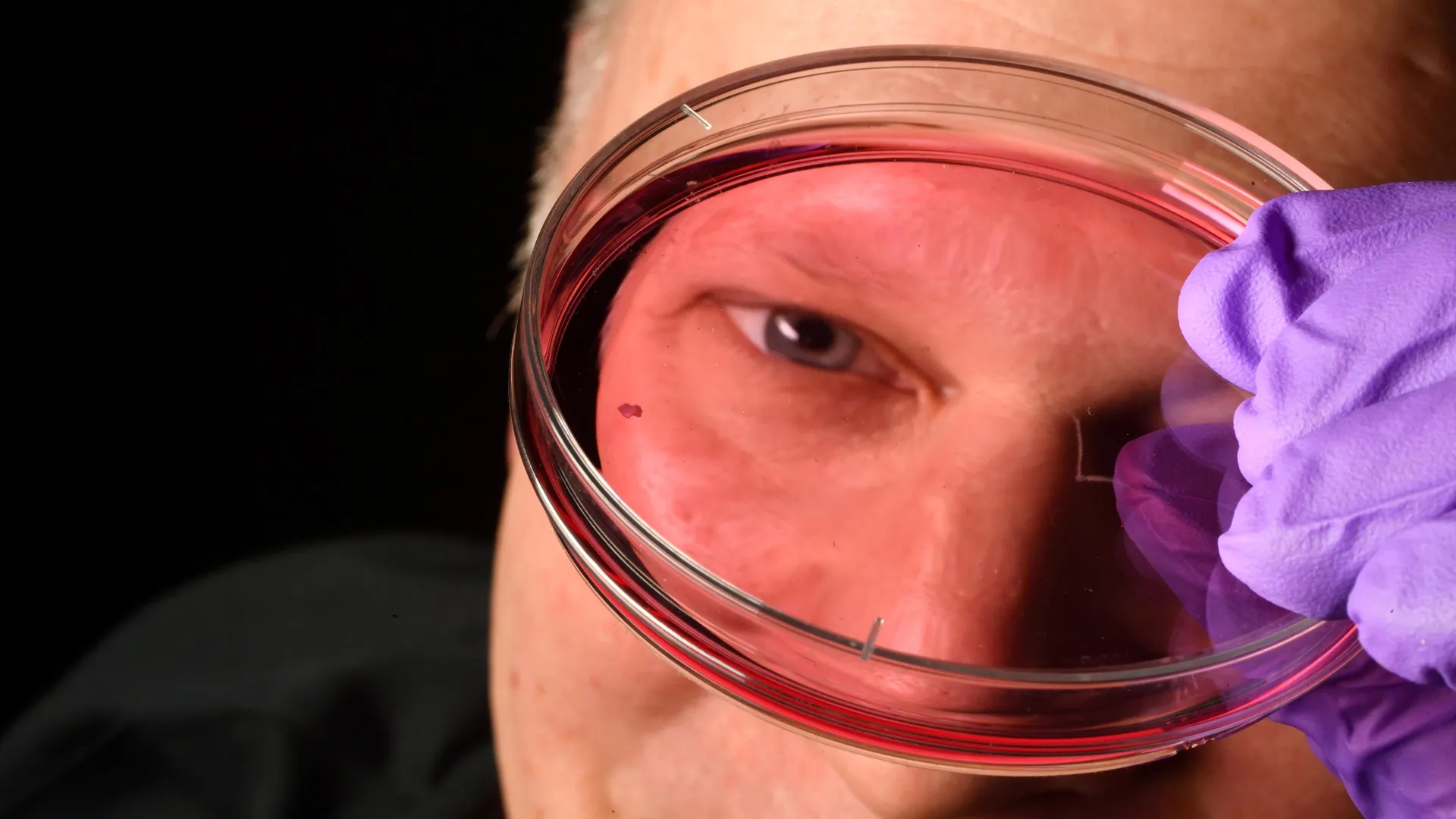
The research team, led by Robert J. Johnston Jr., an associate professor of biology at Johns Hopkins, utilized cutting-edge "organoid" technology to observe the development of the retina in real-time. By cultivating small clusters of tissue from fetal cells, the team was able to create a laboratory-grown model of the human retina that closely mirrors the biological environment of a developing fetus.
Through months of meticulous observation, the researchers identified a precise, two-stage biochemical sequence that occurs between the 10th and 14th weeks of gestation.
Stage 1: The Retinoic Acid Gatekeeper (Weeks 10–12)
During the early stages of fetal development, the retina begins to populate with cone cells. The team discovered that retinoic acid—a molecule derived from vitamin A—acts as a primary regulator. During weeks 10 through 12, retinoic acid signaling is active, facilitating the initial development of blue cones. However, as the developmental window progresses, the concentration of retinoic acid is carefully broken down, which effectively halts the production of new blue-sensing cells.
Stage 2: The Thyroid Hormone Conversion (Weeks 12–14)
The most striking discovery occurred in the following weeks. Rather than migrating away as previously thought, the existing blue cones remain in situ, but undergo a dramatic "identity shift." The researchers identified that thyroid hormones trigger a conversion process, forcing the remaining blue cones to transform into red and green cones.
"First, retinoic acid helps set the pattern," explains Johnston. "Then, thyroid hormone plays a role in converting the leftover cells. That’s very important because if you have those blue cones in there, you don’t see as well. It’s a beautifully coordinated sequence of events."
Challenging the 30-Year Paradigm
For nearly three decades, the prevailing dogma in visual science was one of spatial migration. Textbooks taught that photoreceptors were "born" as specific types and that the arrangement we see in adults was the result of cells shuffling their positions.
Johnston’s team has effectively challenged this static model. By proving that cell identity is fluid during development, they have introduced a dynamic model of retinal formation. "The main model in the field from about 30 years ago was that somehow the few blue cones you get in that region just move out of the way," says Johnston. "We can’t really rule that out entirely, but our data supports a different model. These cells actually convert over time, which is really surprising."
The difficulty in confirming this until now has been the lack of suitable animal models. Common laboratory subjects, such as mice and zebrafish, possess different visual architectures and do not replicate the specific developmental sequence of the human foveola. The use of human-derived retinal organoids was, therefore, the "missing link" that allowed the team to finally observe these processes directly.
Clinical Implications: A New Era for Vision Restoration
The implications of this discovery extend far beyond basic developmental biology. By identifying the exact molecular triggers—vitamin A-derived retinoic acid and thyroid hormones—that dictate cell identity, researchers now have a "chemical key" to manipulate retinal cells in a laboratory setting.
The Quest for "Made-to-Order" Photoreceptors
The ultimate goal of this research is to create cell-replacement therapies for patients suffering from incurable conditions like age-related macular degeneration (AMD). In these patients, the photoreceptors in the central retina die off, leading to a permanent loss of central vision.
If scientists can master the ability to grow healthy, functional photoreceptors in the lab, they could theoretically transplant these cells into a patient’s eye to replace the damaged tissue. "The goal with using this organoid tech is to eventually make an almost made-to-order population of photoreceptors," explains Dr. Sarah Hussey, a former member of the research team and current molecular biologist at CiRC Biosciences.
Bridging the Gap to Clinical Application
While the laboratory results are groundbreaking, the team is careful to manage expectations regarding the timeline for human clinical trials. "These are very long-term experiments," Hussey notes. "Of course, we’d need to do optimizations for safety and efficacy studies prior to moving into the clinic. But it’s a viable journey."
The current focus of the Johnston lab is to further refine the retinal organoids, ensuring they more accurately mimic the physiological complexity of a mature human retina. By doing so, they hope to create a stable, reliable source of photoreceptors that can integrate into existing eye structures without being rejected or failing to function.
The Future of Retinal Medicine
The Johns Hopkins study stands as a testament to the power of organoid technology in modern medicine. By enabling researchers to peer into the "black box" of human fetal development, the team has successfully overturned a 30-year-old scientific consensus.
As the research progresses, the medical community will be watching closely to see if these findings can be scaled from the petri dish to the patient. If the process of cellular conversion—from blue-sensitive to red/green-sensitive—can be reliably replicated in therapeutic contexts, it could provide a permanent, restorative treatment for millions of people currently living in the shadows of vision loss.
The work serves as a reminder that the key to curing the diseases of the future often lies in the most fundamental mysteries of our past—specifically, in the delicate, orchestrated dance of molecules that occurs before we are even born. Through this discovery, the path toward restoring the "sharpness" of human vision has never been clearer.