







For decades, the study of human vascular health has been constrained by a fundamental limitation: the disparity between biological reality and laboratory representation. Human blood vessels are marvels of anatomical complexity—dynamic, twisting, branching, and irregular structures that dictate the fluid dynamics of our life-sustaining circulatory system. Yet, until recently, laboratory models have largely treated these vessels as uniform, straight tubes.
This simplification, while historically useful for basic observation, has left a critical gap in our understanding of how vascular diseases—such as aneurysms and stenosis—actually initiate and progress. A breakthrough from the Department of Biomedical Engineering at Texas A&M University is now closing that gap. Researchers have unveiled a customizable "vessel-chip" system that replicates the intricate architecture of human blood vessels, providing a sophisticated, non-animal platform for testing pharmaceuticals and probing the mechanics of cardiovascular disease.
The Architectural Imperative: Why Shape Matters
The human circulatory system is far from a simple plumbing network. Blood flow is governed by the physical geometry of the vessels through which it travels. When a vessel branches, narrows (stenosis), or suddenly expands (an aneurysm), the physical forces exerted on the vessel walls—known as shear stress—change dramatically.
"There are branched vessels, or aneurysms that have sudden expansion, and then stenosis that restricts the vessel," explains Jennifer Lee, a master’s student in biomedical engineering who led the development of this advanced system. "All these different types of vessels cause the blood flow pattern to be significantly changed, and the inside of the blood vessel is affected by the level of shear stress caused by these flow patterns. That’s what we wanted to model."
By failing to account for these architectural nuances, previous models often missed the very triggers that lead to plaque buildup, clots, and vessel degradation. The new vessel-chip technology, developed in the Bioinspired Translational Microsystems Laboratory, changes the paradigm by allowing researchers to "print" or mold chips that mimic these specific, complex geometries.
Chronology of a Breakthrough
The journey to this innovation began several years ago under the guidance of Dr. Abhishek Jain, an associate professor and the Barbara and Ralph Cox ’53 Faculty Fellow. The evolution of the laboratory’s work highlights a strategic, iterative approach to biomedical engineering.
- The Foundation (Pre-2022): The lab’s early focus centered on establishing the feasibility of organs-on-a-chip technology. Dr. Tanmay Mathur, a former graduate student under Jain, successfully developed a "straight" vessel-chip. This served as the "proof of concept," demonstrating that human cells could be cultured within microfluidic environments to simulate blood flow.
- The Expansion (2023–2024): Jennifer Lee, having joined the lab as an undergraduate honors student, took up the challenge of evolving Mathur’s design. Recognizing that physiological relevance required more than just a tube, Lee worked to incorporate the irregular geometries—bends, branches, and expansions—that define human pathology.
- The Validation (May 2025): The culmination of this research is set to be featured on the cover of the prestigious journal Lab on a Chip in its May 2025 issue. This publication marks a milestone in translating undergraduate curiosity into high-impact, peer-reviewed scientific discovery.
The Science of the "Living" Chip
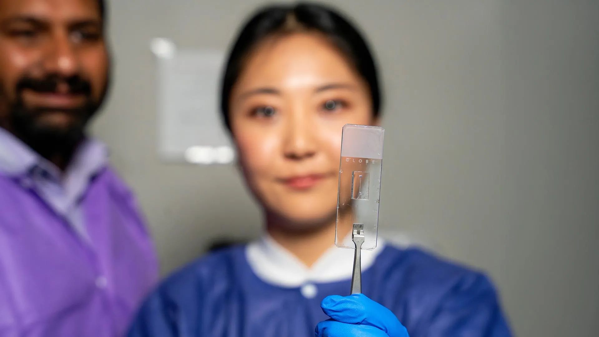
At its core, the vessel-chip is a microfluidic device—a small, transparent platform containing microscopic channels that replicate the dimensions of human blood vessels. However, what sets the Texas A&M model apart is its "living" status.
Unlike inert plastic or silicon models, these chips are seeded with actual human endothelial cells, the specialized cells that line the interior of our blood vessels. When fluid—simulating blood—is pumped through these channels, the cells respond to the mechanical forces of the flow just as they would in a human body.
Dr. Jain emphasizes the importance of this biological realism: "We can now start learning about vascular disease in ways we’ve never been able to before. Not only can you make these structures complex, you can put actual cellular and tissue material inside them and make them living. These are the sites where vascular diseases tend to develop, so understanding them is critical."
Implications for Drug Development and Patient Care
The implications of this technology for the pharmaceutical industry are profound. Currently, drug development relies heavily on animal models, which are often poor predictors of human vascular response, or two-dimensional cell cultures, which lack the mechanical environment of the body.
The vessel-chip offers a "third way"—an in vitro model that is both human-centric and physiologically accurate. This could significantly reduce the time and cost required to bring new cardiovascular drugs to market. Furthermore, because these chips can be tailored to individual patients using their own cells, the technology holds the potential for "personalized medicine." A clinician could theoretically test how a specific patient’s vasculature might react to a drug before prescribing it, effectively minimizing side effects and optimizing treatment efficacy.
Nurturing Scientific Talent: The Undergraduate Path
The success of this project is also a testament to the effectiveness of Texas A&M’s fast-track Master of Science program. Jennifer Lee’s trajectory from a curious undergraduate to a lead researcher on a cover-story project illustrates the university’s commitment to early-career research.
Dr. Jain speaks highly of the academic environment that allowed for this project’s growth. "Jennifer demonstrated perseverance, curiosity, and creativity and started taking up research projects very quickly. Our fast-track program enables students like Jennifer to take on high-impact, high-risk research and not just do a science project, but take it all the way to its outcome and get it published."
For Lee, the experience was about more than just the technical output; it was about the cultivation of a professional mindset. "It’s such a good environment to interact with not only peers but also graduate students and postdoctoral researchers," she says. "You’re able to learn teamwork and communication, work ethic, and just trying different things out. I think it’s such a valuable experience."
Future Frontiers: The Fourth Dimension
As successful as the current model is, the team at the Bioinspired Translational Microsystems Laboratory is already looking toward the next frontier. Currently, the model focuses primarily on endothelial cells. The next iteration aims to introduce multi-cellular complexity. By incorporating smooth muscle cells and other tissue types, researchers hope to observe how different layers of the vessel wall interact with one another under the stress of turbulent blood flow.
Dr. Jain refers to this next step as the "fourth dimensionality" of organs-on-a-chip. "We are progressing and creating what we call the fourth dimensionality of organs-on-a-chip, where we not only focus on the cells and the flow, but this interaction of cells and flow in more complex architectural states, which is a new direction in the field."
A Multi-Institutional Effort
The ambitious scope of this research has garnered support from a vast network of national organizations, underscoring the high stakes and the potential impact of this technology. The research has been funded by a coalition that includes:
- The U.S. Army Medical Research Program
- NASA
- The Biomedical Advanced Research and Development Authority (BARDA)
- The National Institutes of Health (NIH)
- The U.S. Food and Drug Administration (FDA)
- The National Science Foundation (NSF)
- The Texas A&M University Office of Innovation Translational Investment Funds
This widespread interest from government and regulatory bodies suggests that the vessel-chip is viewed not just as a laboratory curiosity, but as a potential standard-bearer for the future of biomedical testing and patient-specific diagnostics.
Conclusion
The work led by Jennifer Lee and Dr. Abhishek Jain at Texas A&M is a poignant reminder that scientific progress often moves forward by refining our understanding of the small. By acknowledging the complex, irregular nature of the human vessel, they have created a tool that could fundamentally alter the landscape of vascular medicine. As the research continues to evolve toward multi-tissue integration, the "fourth dimension" of these chips promises to move us closer to a future where vascular diseases are not only better understood but also more effectively, and perhaps more personally, treated.