











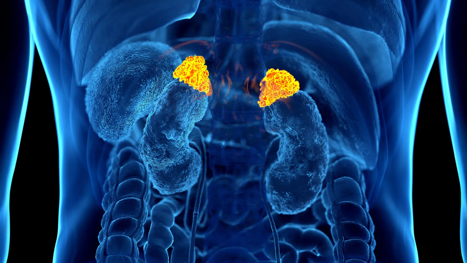
For decades, chronic stress has been described as a "silent killer"—a pervasive, systemic condition that erodes health from the inside out, yet remains notoriously difficult to measure. While clinicians have relied on subjective questionnaires and cumbersome hormonal testing to gauge the impact of stress, a team of researchers from Johns Hopkins University has unveiled a groundbreaking solution. By leveraging deep learning artificial intelligence, scientists have identified the first objective biomarker of chronic stress that can be visualized directly on standard, routine medical imaging.
This revolutionary discovery, set to be unveiled at the upcoming annual meeting of the Radiological Society of North America (RSNA), promises to transform how healthcare providers assess the physiological toll of long-term stress. By repurposing millions of existing chest CT scans, the AI-driven tool provides a window into the cumulative "allostatic load" of a patient’s life, offering a bridge between psychosocial experience and clinical cardiovascular risk.
The Physiological Signature of Stress: A New Paradigm
Chronic stress is far more than a psychological hurdle; it is a biological event. According to the American Psychological Association, the sustained activation of the body’s stress response systems can trigger a cascade of health issues, including anxiety, insomnia, muscular pain, hypertension, and immune suppression. More alarmingly, persistent stress is a known precursor to severe pathologies, including obesity, clinical depression, and heart disease.
Until now, measuring this burden was an imperfect science. Standard methods, such as salivary cortisol testing, offer only a "snapshot" of stress at a specific moment, failing to capture the longitudinal damage accumulated over years. Other methods, such as assessing markers of systemic inflammation, are often non-specific.
The Johns Hopkins team, led by postdoctoral research fellow Elena Ghotbi, M.D., sought a more robust, long-term metric. They focused on the adrenal glands—the body’s "stress command centers." As part of the hypothalamic-pituitary-adrenal (HPA) axis, these glands respond to chronic stress by adjusting their size and activity. The researchers hypothesized that the physical volume of these glands could serve as a permanent, measurable record of an individual’s chronic stress exposure.
Chronology of the Research: From Data to Discovery
The journey to this discovery began by utilizing the Multi-Ethnic Study of Atherosclerosis (MESA), a high-caliber, multi-institutional research initiative that provided the necessary depth to validate such a complex biological marker.
- Data Aggregation: The researchers identified 2,842 participants, with a mean age of 69.3 years, split almost evenly by gender. This cohort was uniquely suited for the study because it contained a "triple-threat" of data: high-resolution chest CT imaging, validated psychosocial stress questionnaires, and comprehensive biochemical markers.
- The Development of the AI Model: Dr. Ghotbi and her team trained a deep learning model to automatically identify, outline, and measure the volume of the adrenal glands in chest CT scans. This automation was critical, as manual measurement would be too time-consuming for clinical practice.
- Defining the Metric: The team established the "Adrenal Volume Index" (AVI), calculated as the total adrenal gland volume (in cubic centimeters) divided by the square of the patient’s height (in meters).
- Validation: To ensure the AVI was a true reflection of stress, the team compared it against salivary cortisol samples taken eight times daily over two days and "allostatic load" scores—a calculation incorporating blood pressure, heart rate, BMI, glucose levels, and other metabolic indicators.
Supporting Data: Validating the Adrenal Volume Index
The results of the study were statistically striking. The AI-derived AVI correlated strongly with multiple indicators of long-term stress. Individuals who reported high levels of perceived stress on standardized questionnaires consistently showed higher AVI values compared to those with lower perceived stress.
Furthermore, the data revealed a clear physiological link between adrenal size and hormonal output. Higher AVI values were directly associated with greater cumulative cortisol exposure and higher peak cortisol levels. Perhaps most significantly, the researchers found a direct link to cardiovascular health.
When observing the participants over a 10-year follow-up period, the team discovered that for every 1 cm³/m² increase in AVI, the risk of heart failure and death increased significantly. The index was also linked to a higher left ventricular mass index—a structural change in the heart often associated with long-term hypertension and cardiac strain. This suggests that the adrenal glands are not merely "responding" to stress; their enlargement is a physical manifestation of a process that is actively damaging the cardiovascular system.
Official Responses and Expert Perspectives
The research has been met with significant enthusiasm within the radiology and cardiology communities, as it addresses a longstanding gap in preventative medicine.
"For the first time, we can ‘see’ the long-term burden of stress inside the body, using a scan that patients already get every day in hospitals across the country," said senior author Shadpour Demehri, M.D., a professor of radiology at Johns Hopkins. Dr. Demehri emphasizes that the utility of this tool lies in its accessibility. Because tens of millions of chest CT scans are performed annually in the United States, the potential for integrating this AI tool into existing diagnostic workflows is vast.
Dr. Teresa E. Seeman, a co-author and professor of epidemiology at UCLA, who has spent three decades studying the impact of stress on the human body, described the breakthrough as a vital step forward. "What makes this work so exciting is that it links a routinely obtained imaging feature with validated biological and psychological measures of stress," Dr. Seeman noted. "It’s a true step forward in operationalizing the cumulative impact of stress on health."
Dr. Ghotbi, the study’s lead author, highlighted the practical advantages for clinical care. "Our approach leverages widely available imaging data and opens the door to large-scale evaluations of the biological impact of chronic stress," Ghotbi stated. "This AI-driven biomarker has the potential to enhance cardiovascular risk stratification and guide preventive care without the need for additional testing or radiation exposure."
Implications: The Future of Preventive Medicine
The implications of this study are profound, particularly for middle-aged and older populations. By quantifying the "allostatic load" of a patient, physicians could potentially identify individuals at high risk for heart failure long before they manifest clinical symptoms.
1. Enhanced Risk Stratification
Currently, heart disease risk is assessed using traditional factors like cholesterol, smoking status, and blood pressure. Adding the Adrenal Volume Index as a "stress metric" could provide a more comprehensive view of a patient’s health profile, accounting for the "invisible" wear and tear caused by lifestyle and psychological stressors.
2. Streamlining Clinical Workflows
Because the AI model is designed to function on scans that are already being performed for other reasons (such as lung cancer screenings or heart evaluations), there is no additional cost to the patient or burden on the healthcare system. The AI acts as a "second set of eyes," extracting diagnostic value from data that would otherwise be ignored.
3. A New Window into Psychosomatic Disease
The ability to map stress to a physical, quantifiable marker opens new avenues for research into a wide array of stress-related illnesses. From autoimmune conditions to cognitive decline, the ability to "see" the physiological consequences of stress could allow researchers to better understand the mechanisms linking the mind and body.
4. Moving Toward Proactive Care
By identifying high AVI in patients, clinicians could move from reactive treatment to proactive intervention. This might include earlier referrals to mental health support, stress-management programs, or more aggressive cardiovascular monitoring—all triggered by a routine imaging scan.
As the medical community prepares to digest these findings at the RSNA annual meeting, the consensus is clear: the era of the "invisible burden" is coming to an end. By turning the lens of artificial intelligence toward the adrenal glands, researchers have provided the medical field with a potent new tool, proving that while stress may be felt in the mind, it leaves a very real, very visible footprint in the body.