










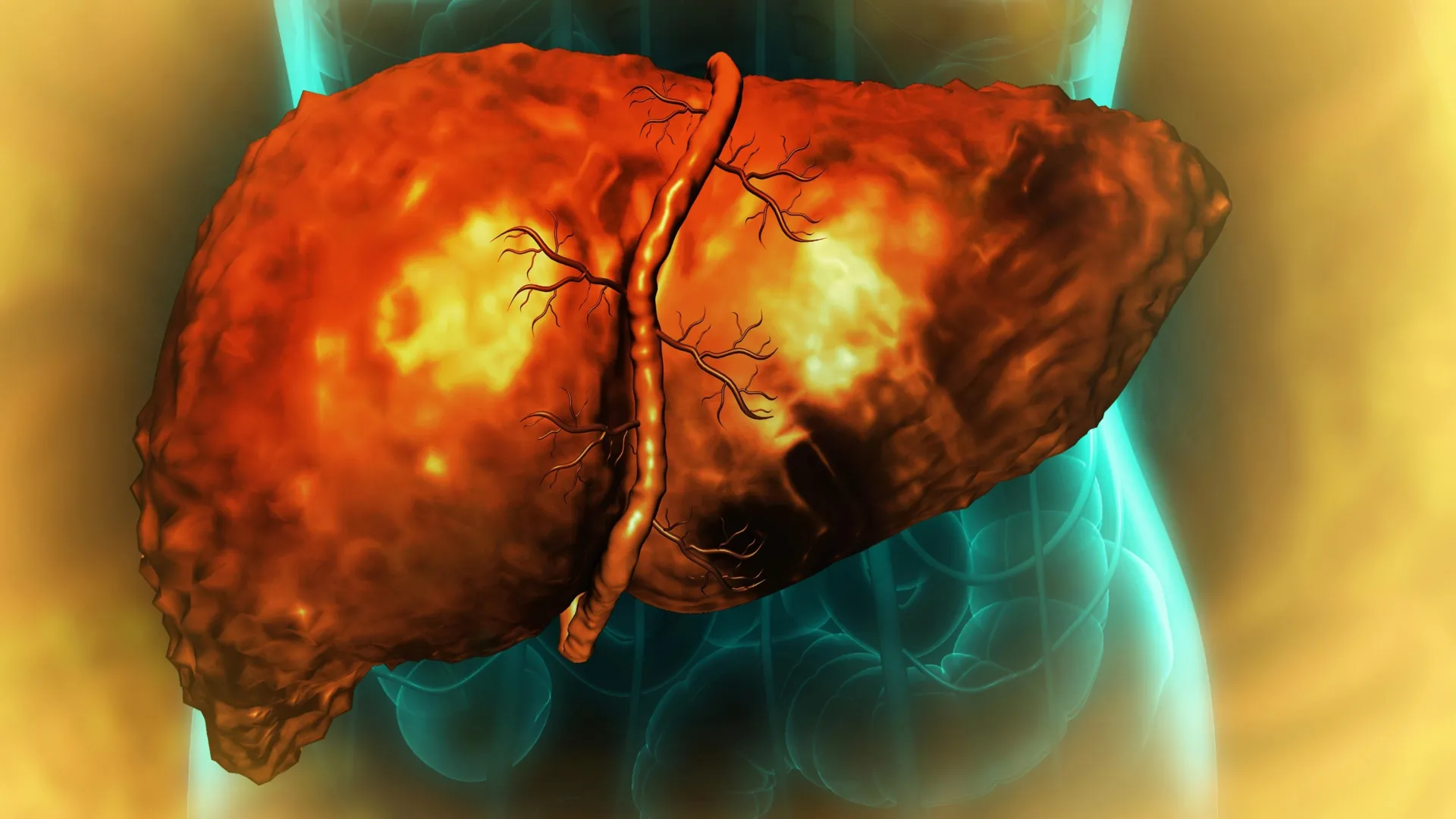
The human liver possesses a biological superpower: the ability to regenerate after trauma. Whether dealing with viral infection, physical injury, or partial surgical removal, this resilient organ typically orchestrates a complex symphony of cellular reprogramming to replace lost tissue. However, in the context of alcohol-associated liver disease (AALD)—a condition responsible for approximately 3 million deaths annually—this regenerative capacity catastrophically fails.
New, groundbreaking research from a multi-institutional team including the University of Illinois Urbana-Champaign, Duke University, and the Chan Zuckerberg Biohub Chicago has finally uncovered why. The study, published in Nature Communications, reveals that chronic alcohol consumption traps liver cells in a "quasi-progenitor" state—a form of cellular limbo where they are neither fully functional nor capable of completing the regenerative cycle. This discovery not only clarifies a long-standing medical mystery but also identifies a specific molecular bottleneck that could pave the way for novel diagnostic markers and life-saving therapeutic interventions.
The Main Facts: A Cycle of Stagnation
The core of the problem lies in the liver’s inability to "finish" the repair process. Under healthy conditions, mature liver cells revert to a fetal-like state, multiply, and then mature back into functioning tissue. In patients with severe alcohol-associated hepatitis or cirrhosis, this cycle is interrupted.
Researchers found that these cells begin the journey toward regeneration but are blocked midway. They are caught in a state of suspended animation, unable to perform their primary metabolic functions or complete the cell division required to replace damaged tissue. As these cells remain trapped, the remaining healthy cells are forced to shoulder an unsustainable workload, leading to increased metabolic stress. This triggers a vicious cycle: healthy cells, pushed to their limit, attempt to regenerate, only to become trapped in the same unproductive state, eventually precipitating total liver failure.
A Chronology of Discovery
The journey to this discovery began with the foundational knowledge that liver cells undergo temporary gene reprogramming during regeneration. Professor Auinash Kalsotra of the University of Illinois and Professor Anna Mae Diehl of Duke University have dedicated years to mapping these molecular processes.
Phase 1: Identifying the "Limbo" State
By comparing healthy liver samples with tissue harvested from patients with alcohol-associated hepatitis and cirrhosis—donated through the National Institute on Alcohol Abuse and Alcoholism—the team identified a consistent, striking pattern. The diseased cells were not dead, nor were they fully functional; they were stuck in an intermediary, unproductive phase.
Phase 2: Uncovering the Role of RNA Splicing
To understand the "why," the researchers looked at the machinery of protein synthesis. They utilized deep RNA sequencing and complex computational analysis to investigate RNA splicing—the essential process of editing genetic "blueprints" before they are translated into proteins. They discovered that thousands of genes were experiencing widespread "mis-splicing" in diseased livers.
Phase 3: The ESRP2 Deficiency
The research team pinpointed a specific protein, ESRP2, as the primary culprit. ESRP2 acts as a gatekeeper, ensuring that RNA is spliced correctly. In the presence of alcohol-related damage, ESRP2 levels plummet. Without this regulator, proteins were not only mis-assembled but were also incorrectly localized within the cell—often getting trapped in the cytoplasm when they were needed in the nucleus to perform regenerative work.
Phase 4: The Inflammation Link
Finally, the researchers traced the reduction of ESRP2 back to systemic inflammation. Alcohol consumption triggers an immune response, flooding the liver with inflammatory and growth factors. These signals directly suppress the production and activity of ESRP2, effectively pulling the emergency brake on the liver’s ability to heal itself.
Supporting Data: The Molecular Architecture of Failure
The precision of this study lies in its shift away from total protein quantification. Many traditional studies measure whether a protein exists in a cell. Kalsotra’s team went further, asking where that protein is and whether it is functional.
Their findings revealed that, in many cases, the quantity of a given protein remained normal, but the protein was useless because it was stuck in the cytoplasm. The "address label" (a sequence within the protein) had been compromised by faulty RNA splicing.
Mouse Model Validation
To solidify these findings, the team utilized mice lacking the gene responsible for ESRP2 production. These mice exhibited the same liver injury patterns and regenerative failure observed in human patients with advanced alcohol-associated hepatitis. This provided a definitive causal link: the loss of ESRP2, driven by inflammation, is a primary driver of the clinical decline seen in human patients.
Official Perspectives and Expert Analysis
The researchers involved in the study emphasize that this discovery changes the paradigm for treating end-stage liver disease.
"We knew that the liver stops functioning and stops regenerating in patients with alcohol-related hepatitis and cirrhosis, even when a patient has discontinued consuming alcohol, but we didn’t know why," said Professor Auinash Kalsotra. "The only real life-saving treatment option once a patient reaches the liver failure stage… is transplantation. But if we understood why these livers were failing, maybe we could intervene."
Graduate students and co-first authors Ullas Chembazhi and Sushant Bangru highlighted the clinical stakes: "They are neither functional adult cells nor proliferative progenitor cells. Since they are not functioning, more pressure builds on the remaining cells. So they try to regenerate, and they’re all ending up in this unproductive quasi-progenitor state, and that’s what is causing liver failure."
The team’s experiment with laboratory cultures further underscored the potential for intervention. By using a molecule to block the inflammatory signaling pathways, the researchers successfully restored ESRP2 levels and corrected the RNA splicing defects, suggesting that the damage might, in theory, be reversible if the inflammatory signal is silenced early enough.
Implications: A Future Beyond Transplantation
The implications of this research are twofold: diagnostic and therapeutic.
Diagnostic Markers
Because mis-spliced RNA molecules are present in high numbers during the early stages of the disease, they could serve as "biological barcodes." Detecting these molecules could allow clinicians to diagnose alcohol-associated liver disease much earlier than is currently possible using standard blood tests or biopsies.
Therapeutic Innovation
The study provides a clear roadmap for drug development. Rather than focusing on complex organ transplants, future treatments could focus on:
- Targeting Inflammation: Utilizing anti-inflammatory agents specifically tuned to block the pathways that suppress ESRP2.
- RNA Repair: Developing therapies that specifically target the splicing machinery to "fix" the genetic instructions before they lead to protein failure.
- Restoring ESRP2: Finding ways to artificially maintain or boost ESRP2 levels in the liver during periods of high alcohol-related stress.
While the path from laboratory to clinical practice is long, this research represents a fundamental pivot. By moving from treating the symptoms of liver failure to addressing the precise molecular "stagnation" of the cells, the medical community may finally be moving toward a day where a failing liver can be repaired rather than replaced.
Research Credits
The multi-institutional effort was made possible by the collaborative work of U. of I. biochemistry graduate students Diptatanu Das and Subhashis Natua; undergraduate students Katelyn Toohill, Ishita Purwar, and Anuprova Bhowmik; Brandon Peiffer and Zhaoli Sun from Johns Hopkins University; Aurelia Leona and Yogesh Goyal from Northwestern University; and Rajesh Dutta from Duke University.
Funding was provided by the National Institutes of Health, the Chan-Zuckerberg Biohub Chicago, the Duke Endowment, and the Muscular Dystrophy Association. As the researchers look toward future clinical studies, the hope remains that these molecular insights will eventually turn the tide on a disease that has historically left clinicians with few options.