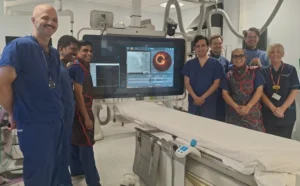


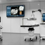


In a significant leap forward for neurodegenerative research, scientists from Nanyang Technological University, Singapore (NTU Singapore) have identified a crucial "clogging" mechanism in the brain that may serve as an early-warning system for Alzheimer’s disease. By analyzing routine magnetic resonance imaging (MRI) scans, researchers have discovered that the brain’s waste-clearance pathways—known as perivascular spaces—often become obstructed long before the onset of clinical dementia symptoms. This discovery offers a potentially transformative tool for clinicians, allowing for earlier intervention in a disease that currently affects tens of millions worldwide.
The Mechanics of Brain Clearance
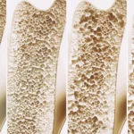
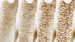
The human brain is an incredibly active organ, constantly generating metabolic waste products such as beta-amyloid and tau proteins. In a healthy brain, these toxic proteins are flushed away through a sophisticated system of perivascular spaces—small, fluid-filled channels that surround blood vessels. When this "drainage system" functions optimally, the brain remains clear of the protein clumps that are the hallmarks of Alzheimer’s disease.
However, the NTU Singapore study reveals that as individuals move toward mild cognitive impairment (MCI), these pathways frequently become enlarged and obstructed. This enlargement acts as a visible marker of systemic failure in the brain’s waste management. Until now, the clinical significance of these enlarged spaces was poorly understood, with many practitioners dismissing them as benign age-related changes. The new research suggests, however, that these "clogged drains" are not merely side effects of aging, but rather a diagnostic window into the underlying pathology of Alzheimer’s.
The Evolution of the Research: A Chronological Overview
The journey to this discovery began with a focus on addressing a massive gap in existing neurological data: the underrepresentation of Asian populations in dementia research. For decades, the global understanding of Alzheimer’s has been shaped primarily by studies involving Caucasian cohorts.
Phase 1: Population-Specific Analysis
Recognizing that dementia manifests differently across ethnic groups, the NTU team initiated a comprehensive study involving nearly 1,000 participants in Singapore. By selecting a demographic that mirrors the multi-ethnic landscape of Singapore, researchers ensured the findings would be broadly applicable to a diverse population. This was essential, as the prevalence of genetic risk factors, such as the apolipoprotein E4 (APOE4) gene, varies drastically between ethnic groups—occurring in up to 60% of Caucasian dementia patients compared to less than 20% in Singaporean patients.
Phase 2: Correlating Scans with Biomarkers
Following the initial participant recruitment, the research team—led by Associate Professor Nagaendran Kandiah of the Lee Kong Chian School of Medicine (LKCMedicine)—conducted a dual-analysis. They compared the visual evidence of enlarged perivascular spaces on MRI scans against blood-based biomarkers. By measuring seven specific biochemicals associated with Alzheimer’s—including tau and beta-amyloid levels—the team sought to establish a definitive link between structural brain changes and molecular pathology.
Phase 3: Longitudinal Benchmarking
The study categorized participants into those with normal cognitive function and those exhibiting signs of mild cognitive impairment (MCI). By tracking the structural integrity of the white matter—the brain’s communication network—alongside the perivascular spaces, the researchers were able to quantify the predictive strength of each marker. The findings revealed that in early stages of decline, enlarged perivascular spaces were a more sensitive indicator of Alzheimer’s-related protein buildup than the traditional white matter damage markers used in clinical settings today.
Supporting Data: Why "Clogged Drains" Matter
The strength of the NTU research lies in its empirical evidence. When the researchers analyzed the relationship between blood markers and structural MRI findings, the data presented a compelling narrative:
- Correlation with Toxicity: Enlarged perivascular spaces were found to be statistically linked to four of the seven tested biochemical markers, signaling a higher concentration of amyloid plaques and tau tangles.
- The Superiority of Early Detection: Among participants with mild cognitive impairment, the correlation between these clogged drainage channels and Alzheimer’s biomarkers was significantly stronger than the correlation with white matter damage.
- Diagnostic Precision: This suggests that relying solely on white matter damage—the current standard—might be missing the "earliest" window of opportunity for medical intervention.
By identifying these anomalies on routine MRI scans, doctors can potentially identify high-risk individuals without the need for expensive, invasive, or highly specialized testing, effectively lowering the barrier to early diagnostic screening.
Official Perspectives and Expert Analysis
The study has sent ripples through the neurological community, garnering praise from experts who emphasize the need to rethink how we diagnose neurodegenerative conditions.
The Clinical Value
Associate Professor Nagaendran Kandiah, Director of the Dementia Research Centre (Singapore) at LKCMedicine, emphasized the practical utility of the findings. "Since these brain anomalies can be visually identified on routine MRI scans performed to evaluate cognitive decline, identifying them could complement existing methods to detect Alzheimer’s earlier, without having to do and pay for additional tests," he stated.
Justin Ong, the study’s first author and a fifth-year medical student at LKCMedicine, highlighted the human impact of these findings. "Identifying Alzheimer’s sooner gives doctors more time to intervene and potentially slow the progression of symptoms such as memory loss, reduced thinking speed, and mood changes," Ong noted.
Bridging the Gap in Neurology
Independent experts have also lauded the study for its contribution to the "synergistic" understanding of brain health. Dr. Chong Yao Feng, a Consultant at the National University Hospital’s Division of Neurology, noted that medicine has historically treated cerebrovascular disease and Alzheimer’s as isolated silos. "The study’s findings are intriguing as they demonstrate that both diseases do interact in a synergistic manner," said Dr. Chong. He cautions, however, that clinicians must exercise judgment: when these markers appear, doctors must look beyond simple vascular issues and consider the possibility of a brewing Alzheimer’s diagnosis.
Dr. Rachel Cheong Chin Yee, a Senior Consultant at Khoo Teck Puat Hospital, echoed these sentiments, noting that the study underscores the importance of vascular health in overall cognitive maintenance. "These findings are significant because they suggest that brain scans showing enlarged perivascular spaces could potentially help identify people at higher risk of Alzheimer’s disease, even before symptoms appear," she remarked.
Implications for Future Care and Treatment
The implications of this research are far-reaching, extending from the clinic to the laboratory and ultimately to the patient’s quality of life.
Redefining Routine Screening
If validated through further longitudinal studies, the observation of perivascular spaces could become a standard feature of neurological reporting. Radiologists and neurologists would be trained to look for these specific structural signatures during standard check-ups, turning routine imaging into a predictive tool for long-term brain health.
Early Intervention Strategies
The goal of modern Alzheimer’s treatment is no longer just symptom management, but disease modification. By catching the disease at the "clogged drain" stage, physicians may be able to implement lifestyle modifications, pharmaceutical interventions, or clinical trial enrollments years before the patient experiences significant loss of independence. This is crucial, as the window for effective therapeutic intervention is often thought to be narrowest once significant, irreversible brain damage has occurred.
A Global Model for Research
The success of the NTU study provides a template for future research in underrepresented populations. By demonstrating that biological markers for Alzheimer’s can behave differently across ethnic groups, the study paves the way for a more personalized, globally representative approach to neurodegenerative medicine.
The Path Forward: What Comes Next
The NTU team is not stopping at these initial findings. Their next phase involves a rigorous longitudinal study, following the participants over an extended period to observe how many eventually transition from mild cognitive impairment to full-scale Alzheimer’s dementia. This data will be instrumental in establishing the predictive power of enlarged perivascular spaces as a reliable clinical tool.
As the global population ages, the burden of dementia will continue to rise. Innovations like those led by the NTU Singapore team offer a beacon of hope, shifting the narrative from a reactive approach—dealing with the devastation of advanced dementia—to a proactive one. By understanding the brain’s waste removal system and identifying the early signs of its failure, science is moving closer to a future where Alzheimer’s can be caught, managed, and perhaps one day, effectively treated long before it takes hold of a person’s life.