


By Sree Roy
The landscape of sleep medicine is undergoing a profound transformation. For decades, the gold standard for diagnosing sleep-disordered breathing was the in-lab polysomnography (PSG) study, where patients were tethered to a suite of sensors in a clinical environment. Central to this process were respiratory inductive plethysmography (RIP) belts—straps placed around the chest and abdomen to measure the rhythmic expansion and contraction of the body. These belts provided the essential data needed to distinguish between obstructive and central breathing events.
However, as diagnostic paradigms shift toward the convenience and accessibility of home sleep testing (HST), the industry is being forced to rethink how respiratory effort is captured, validated, and clinically applied. This evolution is moving beyond simple detection, transforming how we view breathing mechanics, sleep staging, and the longitudinal management of chronic sleep disorders.
The Core Shift: From In-Lab Tradition to Home Innovation
The transition to home-based diagnostics has necessitated a fundamental shift in hardware design and data interpretation. In a controlled lab setting, technicians can adjust belts to ensure optimal signal quality. In the home, however, the patient is often left to manage their own setup. This reality has split the industry into two camps: those simplifying the experience by reducing sensor count, and those doubling down on the precision of dual-sensor technology.
"Breathing during sleep is not just about whether airflow is present or absent," explains Sveinbjorn Hoskuldsson, chief technology officer at Nox Medical. "It is also about whether the patient is making an effort to breathe, how the chest and abdomen are moving, and how those patterns change during events."
As the field moves toward more individualized care, the data derived from RIP is no longer just a secondary metric. It is being leveraged for complex tasks, including sleep staging, arousal detection, and the physiological phenotyping of obstructive sleep apnea (OSA).
Chronology of the RIP Evolution
- The Era of the Gold Standard (1980s–2000s): Respiratory effort measurement becomes synonymous with in-lab PSG, using two RIP belts to capture thoracoabdominal motion.
- The Rise of Portable Monitoring (2010s): Home sleep testing gains traction. Manufacturers begin experimenting with single-belt designs to improve patient comfort and compliance.
- The Innovation Surge (2020–2024): Companies begin introducing "form factor" alternatives, moving away from traditional belts toward adhesive patches and accelerometer-based metrics.
- The Algorithmic Pivot (2025–Present): Advances in AI and signal processing allow for sleep staging and arousal detection directly from respiratory signals, effectively decoupling diagnostic capability from the necessity of EEG.
The Case for Dual-Sensor Precision
For many industry leaders, the decision to maintain a two-belt—or two-sensor—system is not merely a preference; it is a clinical necessity. "Measuring both thoracic and abdominal movement gives a fuller picture of respiratory mechanics than measuring only one compartment," says Hoskuldsson. "The choice to use two belts is about preserving the physiology that matters: respiratory effort, thoracoabdominal coordination, and breathing mechanics."
This redundancy serves as a critical safety net. In home testing, nasal pressure signals—the primary metric for airflow—are notoriously prone to degradation if a patient mouth-breathes or if the cannula becomes displaced. In such instances, the respiratory effort signal becomes the last line of defense for a physician trying to determine if a breathing pause is obstructive (where effort continues despite blocked airflow) or central (where effort is absent).
Amir Reuveny, PhD, CEO of Wesper, echoes this sentiment, noting that the industry has historically compromised on quality for the sake of simplicity. "Many times in sleep medicine, people choose only one sensor because it’s ‘good enough,’" Reuveny says. "But when you move to the home, ‘good enough’ can lead to missed diagnoses."
New Form Factors: Replacing the Belt
One of the most significant barriers to successful home testing has always been the physical discomfort of belts. If a belt is too tight, it interferes with sleep; if it is too loose, it slips or rolls, creating noise in the data. To combat this, several companies are reimagining the hardware entirely.
The Adhesive Revolution
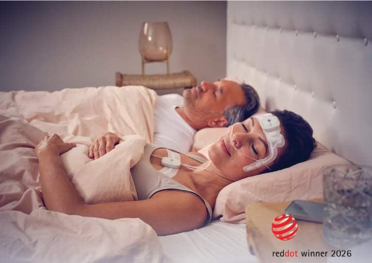
Wesper has pioneered a system that replaces traditional RIP belts with wireless, adhesive patches. These palm-sized, crescent-shaped sensors adhere beneath the breast and over the belly button. According to Reuveny, the design eliminates the "tuning" phase—no tensioning or adjustment is required.
Clinical validation supports this transition. FDA 510(k) K203343, a bench test comparing Wesper’s respiratory effort patches against traditional RIP technology, demonstrated that the adhesive sensors met or exceeded the performance of nasal cannula-utilizing devices across a variety of breathing frequencies, amplitudes, and patient profiles, including those with varying body mass indices and body hair.
The Accelerometer Approach
Huxley Medical has taken a distinct path with its SANSA device, a single-point-of-contact monitor that eliminates belts and patches entirely. Instead of measuring the inductive loop, the device uses an accelerometer-based metric to capture chest wall acceleration and a photoplethysmogram (PPG) to measure respiratory-induced pulse wave modulation.
"Our accelerometer-based effort metric looks at the same thing as a RIP belt; it’s just looking at the acceleration of the chest wall," explains Brennan Torstrick, PhD, chief scientific officer at Huxley Medical. "In addition, we measure the rhythmic changes in blood volume that occur during the ventilatory cycle."
A study presented at SLEEP 2025 showcased the efficacy of this method, reporting 100% sensitivity and 98.9% specificity in identifying central sleep apnea with an index of 15 or more, validating that high-quality data can be captured through non-traditional form factors.
Engineering for Stability: The Case for Better Belts
While some manufacturers move away from belts, others are doubling down on refining them. Research conducted by Nox Medical has highlighted that the design of the belt itself—specifically the material and the integration of contacts—is a primary determinant of signal quality.
"If a belt folds, shifts, stretches inconsistently, or has variable contact quality, the recorded signal becomes less accurate," says Hoskuldsson. "From a clinical perspective, this underscores that the quality of the respiratory signal depends not only on the algorithm, but also on the integrity and consistency of the hardware collecting the data."
General Sleep Corporation has addressed this by eliminating "cut-to-fit" designs, which often suffer from signal loss at secondary connection points. Their reusable belt snaps directly onto the recording unit, ensuring a consistent path for data transmission and mirroring the reliability of in-lab standards.
Clinical Implications: Sleep Staging and OSA Phenotyping
Perhaps the most exciting application of modern RIP technology is its newfound ability to infer sleep stages and arousals without the traditional gold standard of EEG (electroencephalogram). This methodology relies on the well-established physiological link between sleep state and ventilatory control.
Nox Medical’s BodySleep functionality is a prime example of this transition. By analyzing breath-to-breath variability, pattern regularity, and upper airway behavior, the technology can determine whether a patient is in NREM or REM sleep. This is a game-changer for specific patient populations, such as women, who may not exhibit significant oxygen desaturations during respiratory events but whose sleep quality is severely fragmented by cortical arousals.
Furthermore, the industry is increasingly using calibrated dual RIP to perform "physiological phenotyping" of OSA. This allows clinicians to move beyond a simple Apnea-Hypopnea Index (AHI) number to understand the why behind a patient’s condition. Is it due to airway collapsibility, a low arousal threshold, or ventilatory control instability? Such insights are essential for moving toward precision medicine, where therapy is tailored to the underlying mechanism of the disorder rather than a generic diagnosis.
Future Directions: Longitudinal Monitoring
The current state of sleep medicine remains heavily focused on the diagnostic snapshot—a single night of testing to confirm a disorder. However, the future of respiratory effort monitoring lies in longitudinal tracking.
"The respiratory effort measurements we provide allow physicians to monitor central apnea over time," says Wesper’s Reuveny. "It can be either therapy-induced or emergent."
As hardware becomes less intrusive and data analytics more robust, the potential to monitor patients throughout their treatment journey becomes a reality. If a therapy—whether it be CPAP, oral appliances, or nerve stimulation—changes the patient’s upper airway behavior or ventilatory stability, these shifts can be tracked and quantified.
The shift to home testing is not just a trend; it is a fundamental reconfiguration of the diagnostic toolkit. By bridging the gap between the controlled environment of the lab and the natural environment of the home, the latest advancements in RIP technology are ensuring that "physiologic truth" is maintained, no matter where the patient sleeps. As these technologies continue to mature, the focus remains clear: providing clinicians with the granular data necessary to improve outcomes, enhance patient comfort, and bring precision sleep medicine into the home.
References
- US Food & Drug Administration. Re: K203343. 2021 Dec 21.
- Khayat R, et al. Detecting central sleep apnea using a multi-diagnostic chest-worn monitor. Sleep. 2025;48(suppl1):A185.
- Montazeri K, et al. The design of RIP belts impacts the reliability and quality of the measured respiratory signals. Sleep Breath. 2021;25(3):1535-41.
- US Food & Drug Administration. Re: K252330. 2025 Nov 17.
- Finnsson E, et al. Detecting arousals and sleep from respiratory inductance plethysmography. Sleep Breath. 2025;29(2):155.
- US Food & Drug Administration. Re: K241960. 2025 Mar 14.
- Finnsson E, et al. Respiratory inductance plethysmography to quantify changes in ventilation in obstructive sleep apnea. IEEE Trans Biomed Eng. 2026;73(5):1943-52.