





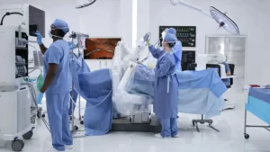

For decades, the field of cardiology has operated under a sobering, fundamental limitation: the human heart is largely incapable of self-repair. Unlike skin or bone, which possess robust regenerative properties, the adult heart responds to injury—whether from a myocardial infarction (heart attack), chronic hypertension, or viral myocarditis—by replacing healthy, contractile muscle cells with non-functional, rigid scar tissue. This process, known as cardiac remodeling, inevitably leads to heart failure, a progressive condition where the organ struggles to circulate blood effectively throughout the body.
While the modern pharmacological arsenal has expanded significantly—with recent breakthroughs such as GLP-1 receptor agonists (obesity drugs) showing promise in reducing heart failure symptoms—these treatments remain largely palliative. They manage the disease rather than reversing the underlying structural damage. For many patients, the end of the road is a binary choice: a high-risk, donor-dependent heart transplant or the implantation of a mechanical left ventricular assist device (LVAD).
Now, a pioneering new study published in the New England Journal of Medicine offers a glimpse into a third, transformative path: the surgical implantation of lab-grown, engineered heart muscle patches.
The Science of BioVAT: A New Frontier in Cardiology
The recent study highlights the successful clinical application of a novel therapeutic, dubbed "BioVAT" (Biological Ventricular Assist Tissue). These patches are constructed from induced pluripotent stem cells (iPSCs)—adult cells that have been reprogrammed into a stem-like state, allowing them to be coaxed into becoming functional cardiac muscle cells (cardiomyocytes).
Unlike traditional cellular therapies, which often involve injecting isolated cells into the heart—a method plagued by poor cell retention and the risk of arrhythmias—BioVAT is an organized, three-dimensional tissue construct. By engineering these cells into a structured patch, researchers provide the damaged heart with a "scaffold" that mimics the architectural integrity of natural myocardium. Once sutured onto the epicardium of the damaged heart wall, these patches integrate with the patient’s existing tissue, theoretically providing a functional layer of contractile muscle that "revs up" the heart’s weakened pumping capacity.
A Chronology of Cardiac Repair
The journey to this clinical milestone has been marked by decades of laboratory trial and error.
- Early 2000s: The discovery of iPSCs by Shinya Yamanaka revolutionized regenerative medicine. For the first time, researchers had a scalable, ethically non-controversial source of human cells capable of becoming any tissue type.
- 2010–2015: Preclinical studies in rodent and porcine models demonstrated that cardiomyocytes derived from stem cells could indeed beat in synchrony with host tissue. However, the hurdle of "electromechanical integration"—ensuring the new cells beat in time with the rest of the heart—remained a significant barrier.
- 2018–2022: The development of sophisticated bio-scaffolds allowed researchers to create "patches" rather than loose cell suspensions. This period saw the refinement of manufacturing protocols to ensure that these patches were not only contractile but also durable enough to survive the mechanical stress of a beating human heart.
- 2024–2025: The first clinical application of BioVAT in patients with end-stage heart failure. The recent study results represent the first time this technology has demonstrated clear, measurable improvement in human heart wall thickness and pumping efficiency in a clinical setting.
Supporting Data: Translating Promise into Metrics
The preliminary data from the new study provides a compelling case for the efficacy of BioVAT. In the cohort of patients who received the engineered patches, researchers observed three primary clinical signals:

- Structural Re-muscularization: Post-operative imaging, including advanced MRI and echocardiography, revealed a measurable thickening of the ventricular walls at the site of the patch application. This suggests that the engineered tissue successfully integrated and began to function as a supplementary muscle layer.
- Improved Ejection Fraction: The primary metric of heart performance—the Left Ventricular Ejection Fraction (LVEF)—showed a modest but statistically significant improvement. This increase in the percentage of blood pumped out of the heart per beat translates directly to improved perfusion of vital organs.
- Functional Capacity: Patients reported a quantifiable improvement in their quality of life, including increased exercise tolerance and a reduction in the debilitating fatigue and shortness of breath that characterize late-stage heart failure.
While these results are preliminary, they suggest that BioVAT may function effectively as a "bridge to recovery" or a "bridge to decision." By bolstering the heart’s function, it may buy patients critical time while they wait for a donor organ or allow them to avoid the complications often associated with long-term mechanical LVAD support, such as infection or stroke.
Official Perspectives and Expert Caution
The medical community has reacted with cautious optimism. Dr. Elena Rodriguez, a regenerative medicine specialist not involved in the study, notes that while the data is "a landmark achievement," the field must remain grounded in the reality of the challenges ahead.
"The leap from a small-scale study to a broader clinical practice is immense," Dr. Rodriguez stated. "We need to understand the long-term immunological response to these patches. Even though they are derived from pluripotent cells, the process of tissue engineering introduces variables that could trigger inflammation or immune rejection over time."
Furthermore, regulatory bodies, including the FDA, have signaled that the path to approval for such "living therapies" will be rigorous. The manufacturing complexity—creating custom, patient-specific heart tissue—presents a logistical hurdle that standard pharmaceutical manufacturing does not face.
"We are essentially talking about moving from ‘off-the-shelf’ pills to ‘bespoke’ biological organs," said a representative from a leading cardiovascular research institute. "The validation of the manufacturing consistency is as important as the clinical outcomes themselves."
Implications for the Future of Heart Failure Care
The implications of successful BioVAT therapy are profound. If the technology proves durable and scalable, it could fundamentally disrupt the current treatment algorithm for heart failure.
1. Reducing the Transplant Waiting List
The most immediate benefit would be the reduction of the "transplant gap." With thousands of patients waiting for hearts that never arrive, any therapy that improves the status of a patient—either by stabilizing them or by potentially recovering enough function to remove them from the transplant list—is a life-saving intervention.

2. Personalized Medicine
Because these patches can be generated from a patient’s own iPSCs, the risk of rejection is theoretically minimized. This personalized approach to cardiac surgery could usher in an era where cardiovascular surgeons act as "biological architects," tailoring the repair to the specific geography of a patient’s injury.
3. A Shift in Therapeutic Strategy
If we can treat the heart as a regenerative organ rather than a static mechanical pump, our goal shifts from maintenance to restoration. This would necessitate a massive increase in funding and research into bioengineering, as well as a new framework for how insurance providers and health systems value "living" surgical interventions compared to traditional medical devices.
The Path Forward: Scaling the Solution
The upcoming larger trials will be the true test for BioVAT. These studies are designed to address the most critical questions remaining:
- Durability: Do the patches remain viable and functional five or ten years post-implantation?
- Patient Selection: Which subset of heart failure patients—ischemic vs. non-ischemic—stands to benefit the most?
- Cost-Benefit Analysis: Given the intensive labor required to engineer this tissue, how can the cost be managed to ensure equitable access across health systems?
As the study moves into its next phase, the medical community remains hopeful. The heart, long thought to be a one-way street toward decline, may finally be entering an era where it can be rebuilt. For the millions of people living in the shadow of heart failure, this research represents more than just data; it represents the hope that the heart’s rhythm, once damaged, might one day be restored.
While we are not yet at a point where a "patch-and-go" solution is standard practice in every community hospital, the bridge from the laboratory to the bedside has been firmly established. The next few years of clinical trial data will determine whether BioVAT becomes the cornerstone of a new cardiac revolution or a stepping stone toward even more sophisticated, perhaps even fully bioprinted, organ replacement therapies. For now, the prospect of cardiac regeneration is no longer a matter of "if," but "when."