










For decades, the standard approach to understanding human vascular biology in a laboratory setting has been defined by a fundamental simplification: the straight, uniform tube. While these simplified models have served as the bedrock of biomedical research, they suffer from a glaring disconnect from physiological reality. Human blood vessels are rarely linear conduits; they are dynamic, branching, narrowing, and widening networks that dictate the fluid mechanics of the circulatory system.
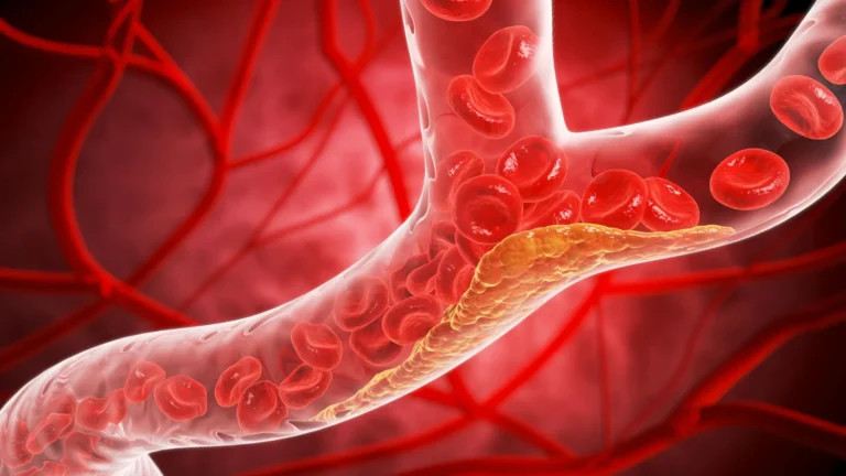
When researchers treat vessels as uniform pipes, they ignore the very geometries where pathology takes root. Aneurysms, stenoses, and complex bifurcations create chaotic flow patterns that fundamentally alter the shear stress experienced by the vessel wall. Recognizing this gap, a team at the Texas A&M University Department of Biomedical Engineering has unveiled a transformative "vessel-chip" system. This customizable microfluidic platform promises to bridge the divide between simplistic benchtop models and the complex, living reality of human vascular health.
The Genesis of Complexity: A Shift in Paradigms
The human circulatory system is a masterpiece of fluid dynamics, yet our ability to model it has historically been hindered by the limitations of manufacturing technology. Laboratory-grown vessels have long been limited to rigid, straight-line configurations. However, vascular diseases—such as atherosclerosis, which often develops at branching points where blood flow becomes turbulent—do not occur in straight pipes.
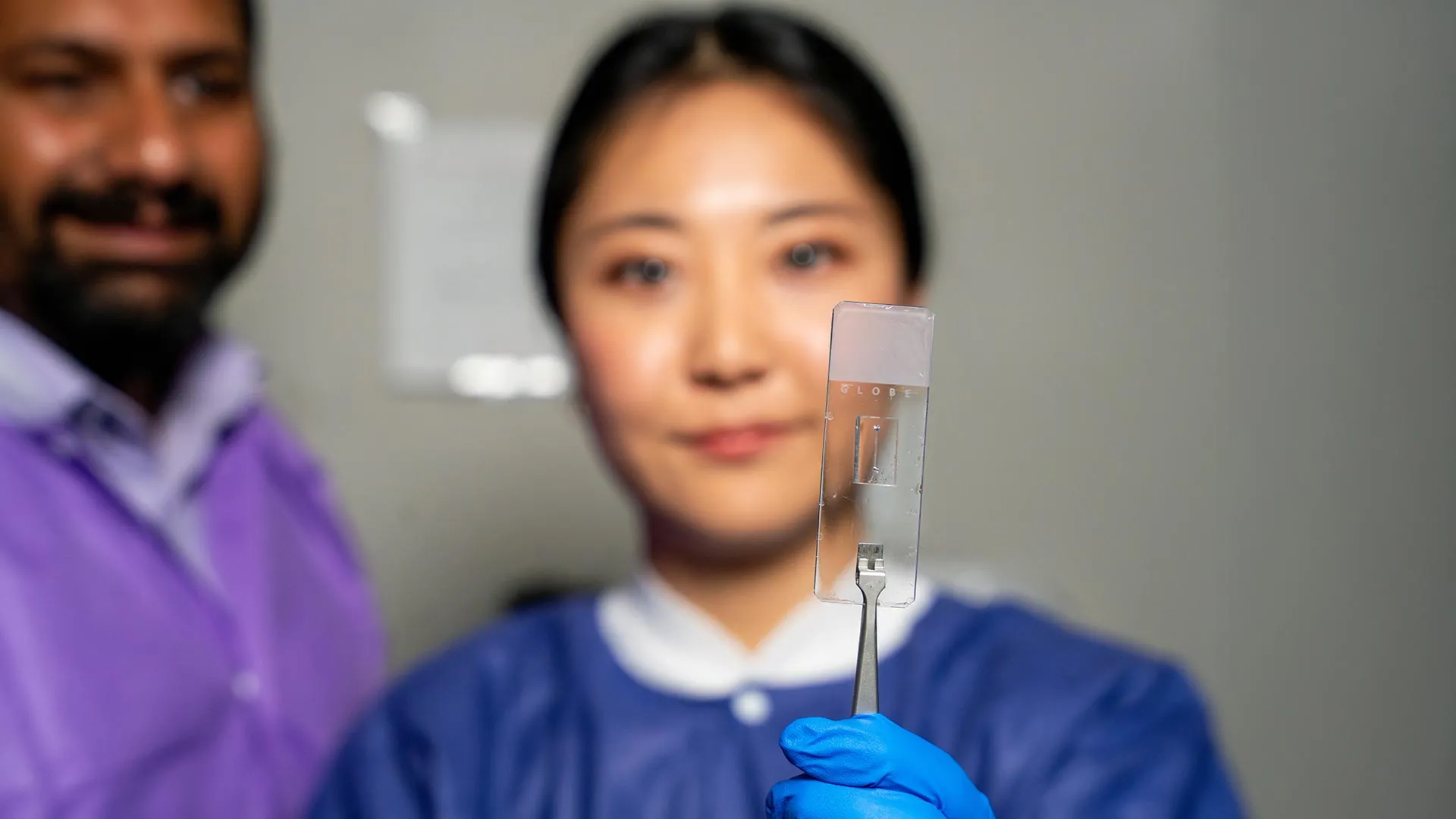
The new vessel-chip system, developed in the Bioinspired Translational Microsystems Laboratory under the guidance of Dr. Abhishek Jain, represents a departure from this "straight-line" orthodoxy. By utilizing advanced microfluidic manufacturing, the team has created a platform capable of replicating the precise, irregular geometries of human vasculature.
Jennifer Lee, a master’s student in biomedical engineering and the lead researcher on the project, explains the motivation behind the leap in design: "There are branched vessels, or aneurysms that have sudden expansion, and then stenosis that restricts the vessel. All these different types of vessels cause the blood flow pattern to be significantly changed, and the inside of the blood vessel is affected by the level of shear stress caused by these flow patterns. That’s what we wanted to model."
By capturing these nuances, the team is effectively moving the study of vascular disease from the realm of the theoretical into a space that mirrors the patient’s actual anatomy.
Chronology: Building on a Foundation of Innovation
The path to the current breakthrough was not an overnight success but a deliberate, multi-year evolution of laboratory methodology.
The Foundational Phase
The current research builds upon the work of Dr. Tanmay Mathur, a former graduate student in the Jain lab. A few years ago, Mathur pioneered the initial straight-vessel-chip design. While seemingly simple in retrospect, that project established the necessary protocols for seeding living tissue into microfluidic environments and managing fluid dynamics at the micron scale.
The Innovation Phase
Jennifer Lee joined the lab as an undergraduate honors student, initially with limited exposure to organs-on-a-chip technology. Her trajectory exemplifies the "fast-track" philosophy of the department. Under Dr. Jain’s mentorship, Lee rapidly moved from foundational learning to independent, high-stakes experimentation. Her focus shifted from replicating simple tubes to engineering the complex, patient-specific architectures that characterize the new vessel-chip.
The Publication Phase
The rigor of this research culminated in a feature in the journal Lab on a Chip. The study, which documents the methodology and validation of these complex, living vessel structures, has been selected for the cover of the journal’s May 2025 issue, signaling its high level of impact within the global biomedical engineering community.
Supporting Data: Why Geometry Matters
To understand the necessity of this technology, one must look at the physics of blood flow. In a straight, uniform vessel, blood typically flows in a laminar, predictable fashion. However, when a vessel narrows (stenosis) or widens (aneurysm), the fluid dynamics change dramatically.
- Shear Stress Variations: The "vessel-chip" allows researchers to measure how cells respond to varied shear stress. High or erratic shear stress is a known precursor to inflammation and the development of plaques.
- Cellular Responsiveness: The current iteration of the chip incorporates endothelial cells—the critical, thin layer of cells that line the interior of every blood vessel. Because these cells act as sensors for blood flow, the chip provides a "living" readout of how vessel geometry affects biological health.
- Non-Animal Alternatives: By providing a highly accurate model of human physiology, the platform offers a path to reduce reliance on animal testing. This not only aligns with ethical advancements in research but also provides more relevant data, as animal vascular systems often differ significantly from human counterparts in their response to drugs.
Official Responses: A New Frontier in "Fourth-Dimensional" Research
Dr. Abhishek Jain, an associate professor and the Barbara and Ralph Cox ’53 faculty fellow in biomedical engineering, views this development as a foundational step toward a new era of personalized medicine.
"We can now start learning about vascular disease in ways we’ve never been able to before," Dr. Jain remarked. "Not only can you make these structures complex, you can put actual cellular and tissue material inside them and make them living. These are the sites where vascular diseases tend to develop, so understanding them is critical."
The lab’s vision extends beyond the current iteration. According to Dr. Jain, the team is working toward what he terms the "fourth dimensionality" of organs-on-a-chip technology.
"We are progressing and creating what we call the fourth dimensionality of organs-on-a-chip, where we not only focus on the cells and the flow, but this interaction of cells and flow in more complex architectural states," Jain explained. By adding further cell types—such as smooth muscle cells or immune cells—the researchers intend to create a multi-layered, interactive tissue model that can simulate the progression of chronic diseases in real-time.
Implications for Clinical Medicine and Pharmacology
The implications of this technology are vast, touching upon both basic scientific understanding and high-stakes clinical applications.
1. Drug Development and Testing
Currently, the process of drug discovery is plagued by high failure rates in clinical trials. A significant portion of these failures occurs because drugs that show promise in simple petri dishes or animal models fail to account for the complex mechanical environment of the human vascular wall. By using patient-specific vessel-chips, pharmaceutical companies could test the efficacy and toxicity of new drugs on "living" human tissue that mimics the specific disease states of their target demographic.
2. Personalized Medicine
Because these chips can be tailored to individual patients, they offer the potential for "clinical trials in a chip." Doctors could theoretically take cells from a patient, seed them into a chip that mimics the patient’s own vascular architecture, and test which treatments are most effective before administering them to the patient. This transition from "one-size-fits-all" to precision cardiology could save countless lives and significantly reduce healthcare costs.
3. Training the Next Generation of Scientists
The project also serves as a case study for academic excellence. Jennifer Lee’s experience underscores the value of Texas A&M’s fast-track research program. By integrating undergraduate and master’s students into high-impact research early in their careers, the department is cultivating a workforce capable of tackling the most difficult challenges in modern medicine.
As Lee notes, the experience provided more than just technical data. "It’s such a good environment to interact with not only peers but also graduate students and postdoctoral researchers," she said. "You’re able to learn teamwork and communication, work ethic, and just trying different things out."
Collaborative Support and Future Outlook
The success of the vessel-chip project is a testament to the power of interdisciplinary collaboration and substantial institutional support. The research has been bolstered by a coalition of high-level partners, including the U.S. Army Medical Research Program, NASA, the Biomedical Advanced Research and Development Authority (BARDA), the National Institutes of Health (NIH), the U.S. Food and Drug Administration (FDA), the National Science Foundation (NSF), and the Texas A&M University Office of Innovation Translational Investment Funds.
As the team prepares for the May 2025 journal release, the horizon for this technology remains bright. By continuing to add layers of biological complexity—such as immune system interaction and metabolic processes—the Bioinspired Translational Microsystems Laboratory is positioning itself at the absolute forefront of cardiovascular research.
The days of viewing the vascular system as a simple, static tube are coming to an end. Through the work of researchers like Jennifer Lee and Dr. Abhishek Jain, the scientific community is finally gaining the tools to view the human body with the complexity and dynamism it deserves. The "living" vessel-chip is not just a device; it is a mirror reflecting the intricate, branching reality of human life, and a vital key to unlocking the future of medicine.