


Chronic ischemic gastritis (CIG) remains one of the most challenging diagnoses in modern gastroenterology. Often masquerading as common peptic ulcer disease or refractory dyspepsia, this vascular pathology is characterized by a significant reduction in blood flow to the stomach, typically secondary to mesenteric artery disease. When left untreated, the consequences are catastrophic, with thromboembolic events posing a direct threat to patient life.
A recent clinical case report published in the medical literature has shed new light on this elusive condition. By identifying a specific, progressive endoscopic marker—the "white patch"—researchers have provided clinicians with a potential diagnostic roadmap that could move beyond the limitations of standard tissue biopsies and patient history.
Main Facts: The Clinical Challenge of CIG
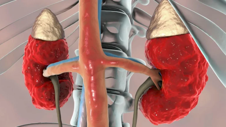
Chronic ischemic gastritis occurs when the arterial supply to the stomach—derived from the celiac trunk and the superior mesenteric artery (SMA)—becomes compromised. Unlike acute ischemia, which presents with sudden, severe pain and systemic shock, chronic ischemia manifests as a smoldering, progressive condition.
The diagnostic dilemma lies in the lack of pathognomonic symptoms. Patients frequently report non-specific epigastric pain, weight loss, and recurring ulcers that fail to respond to traditional proton pump inhibitor (PPI) therapy. Standard diagnostic protocols, such as upper endoscopy and mucosal biopsy, are frequently inconclusive. While endoscopy can visualize the state of the gastric lining, it often fails to pinpoint the underlying vascular etiology, leading to repeated, ineffective treatments for suspected acid-peptic disease.
The core of the recent breakthrough is the identification of a morphological pattern: the evolution of "white patches" on the gastric mucosa. This finding serves as a potential "canary in the coal mine," signaling that the gastric tissue is failing due to hypoperfusion, thereby triggering the need for high-resolution vascular imaging, such as computed tomography (CT) angiography.
Chronology of a Case Study
The significance of these findings is best illustrated through the case of a 63-year-old male patient, whose journey highlights the diagnostic hurdles inherent in CIG.
The Initial Presentation
The patient arrived at the clinic complaining of chronic, persistent epigastric pain. His medical history was marked by a series of recurrent gastric ulcers that had been managed conservatively at various medical facilities. Despite multiple rounds of treatment, the ulcers remained unexplained, and the patient’s pain continued to cycle, significantly impacting his quality of life.
The Endoscopic Turning Point
During the initial workup, the medical team performed a high-definition upper endoscopy. While previous examinations had noted standard inflammatory changes, the current procedure revealed a distinct and evolving pattern: the presence of multiple white patches scattered across the gastric mucosa. These were not typical ulcerations but rather areas of pallor, suggesting ischemic compromise rather than inflammatory erosion.
Vascular Investigation
Recognizing that these patches were inconsistent with standard gastritis, the clinical team shifted their focus from the gastric lumen to the arterial supply. A CT scan of the abdomen was ordered, specifically targeting the mesenteric vasculature. The imaging confirmed a dire vascular situation: complete occlusion of the superior mesenteric artery and significant stenosis of the celiac artery. The "white patches" were, in fact, the visible markers of the stomach’s struggle to receive oxygenated blood.
Intervention and Recovery
Following the diagnosis, the patient underwent a surgical bypass procedure to restore blood flow to the digestive tract. The outcome was transformative. Post-operative follow-ups showed a complete resolution of the white mucosal patches and a total cessation of the patient’s chronic abdominal pain. This success underscored that the gastric mucosa acted as a reliable indicator of the systemic vascular health of the abdomen.
Supporting Data: The Anatomy of Ischemia
The stomach possesses a rich collateral blood supply, which is both a blessing and a diagnostic curse. The network formed by the celiac axis and the SMA ensures that the organ is highly resilient. However, this resilience often masks the onset of ischemia until the stenosis becomes critical.
The Role of Computed Tomography (CT)
While endoscopy provides the visual "clue," CT angiography remains the gold standard for confirmation. Data suggests that in cases of suspected chronic mesenteric ischemia, CT imaging can accurately visualize the degree of luminal narrowing. The recent case emphasizes that clinicians should maintain a low threshold for ordering CT scans when endoscopic findings appear "atypical" or when ulcers do not heal despite optimal medical therapy.
The "White Patch" Phenomenon
Pathophysiologically, the white patches represent mucosal areas experiencing microvascular failure. As blood flow drops, the epithelial cells in the gastric lining undergo metabolic stress, leading to a loss of the vibrant, pink coloration associated with healthy, well-perfused tissue. These white patches represent the transition from reversible ischemia to localized tissue necrosis. Recognizing these patches as a precursor to serious thromboembolic events is a critical advancement in preventative gastroenterology.
Official Responses and Clinical Perspectives
The medical community has long recognized that chronic ischemia is under-diagnosed. In response to this case, gastrointestinal experts have emphasized the need for a more integrated approach to abdominal pain.
"We often treat the stomach as a standalone organ," notes one leading gastroenterologist. "However, this case serves as a vital reminder that the gastric mucosa is a mirror for the entire splanchnic circulation. When we see recurring, unexplained ulcers or strange mucosal changes that don’t fit the ‘H. pylori’ or ‘NSAID’ narrative, we must look at the vasculature."
Many academic medical centers are now proposing that "gastric ischemia" be included in the differential diagnosis for any patient over the age of 60 presenting with refractory epigastric pain. There is a growing consensus that endoscopic reports should explicitly document the appearance and distribution of such white patches, as these could eventually be codified into a formal classification system to assist less experienced clinicians in identifying the condition earlier.
Implications for Future Practice
The implications of these findings are profound for both patient care and medical education.
Enhanced Diagnostic Protocols
The most immediate implication is the potential for a change in diagnostic algorithms. Currently, when an endoscopy is performed, the focus is largely on identifying infection, inflammation, or malignancy. By adding "ischemic markers" to the training curriculum for gastroenterologists, the medical field could significantly reduce the time between symptom onset and surgical intervention.
Patient Outcomes and Mortality
CIG is not merely a quality-of-life issue; it is a life-threatening condition. The risk of sudden mesenteric infarction is high if the underlying stenosis is ignored. By leveraging endoscopic findings to trigger vascular imaging, clinicians can intervene before a fatal thromboembolic event occurs. The case of the 63-year-old patient proves that bypass surgery can reverse the damage, provided the diagnosis is made while the tissue remains salvageable.
Future Research Directions
The identification of white patches opens the door for further research. Future studies should aim to determine if these patches correlate with specific degrees of arterial stenosis or if they appear only after a certain threshold of blood flow reduction is crossed. Furthermore, there is a need to determine if pharmacological management—such as antiplatelet therapy or aggressive lipid-lowering treatments—can stabilize these lesions in patients who are not candidates for major vascular surgery.
Conclusion: A Shift in Paradigm
The story of this 63-year-old patient is more than just a single successful surgery; it is a blueprint for future diagnostic success. By looking beyond the obvious signs of ulcers and paying attention to the subtle, visual cues of the gastric mucosa, physicians can save lives. As we move forward, the "white patch" may well become a standard diagnostic landmark, moving us one step closer to solving the mystery of chronic ischemic gastritis and ensuring that patients receive the surgical intervention they need before it is too late. The stomach, it appears, has been telling us its secrets all along; it is time for clinicians to learn the language.