










November 4, 2026 — By Medical News Correspondent
In the ongoing quest to eradicate multiple myeloma, clinicians have long grappled with a significant hurdle: the limitations of conventional imaging. While treatment protocols have evolved significantly over the last two decades, our ability to visualize the full extent of the disease burden has remained stubbornly suboptimal. A landmark phase II clinical trial, the results of which were published this week in Radiology, suggests that a novel CD38-targeted immuno-PET tracer may finally provide the clarity oncologists have been seeking.
The Challenge of Suboptimal Detection
Multiple myeloma, a cancer of the plasma cells, frequently leaves patients in a precarious diagnostic limbo. The current standard-of-care—fluorine 18 (18F)-fluorodeoxyglucose (FDG) PET/CT—relies on measuring the glucose uptake of rapidly dividing cells. While effective in many cancers, this method is fundamentally limited in the context of plasma cell disorders. Myeloma cells often exhibit low glucose metabolism, leading to a high rate of "false negatives."
Research indicates that as many as 30% to 50% of patients with established bone lesions may have disease that remains entirely undetected by standard FDG PET/CT. This diagnostic gap means that oncologists are often forced to make treatment decisions based on incomplete maps of the patient’s actual disease burden.
The Innovation: CD38-Targeted Immuno-PET
The study, led by Gary A. Ulaner, MD, PhD, of the University of Southern California, explored the efficacy of a new tracer: 89Zr-DFO-daratumumab. This approach represents a shift toward "immuno-PET," a technology that bridges the gap between targeted immunotherapy and molecular imaging.
By utilizing daratumumab—a monoclonal antibody already established as a potent treatment for multiple myeloma—researchers created a tracer that specifically binds to the CD38 protein, which is highly expressed on the surface of myeloma cells. When labeled with the radioisotope Zirconium-89 (89Zr), this tracer acts as a homing beacon, allowing for high-resolution, whole-body mapping of tumor cells, regardless of their glucose metabolism.
Chronology of the Clinical Trial
The prospective, two-center trial was conducted over a three-year period, from March 2022 to July 2025. The study enrolled 60 participants with a median age of 63, all of whom had pathologically proven CD38-positive multiple myeloma.
To ensure the validity of the data, the inclusion criteria were rigorous: participants were required to have at least one lesion previously identified via conventional imaging (CT, MRI, or FDG PET/CT) within 90 days of enrollment. Furthermore, all participants were candidates for either first-line or subsequent lines of myeloma therapy, ensuring the findings would be directly applicable to active clinical care.
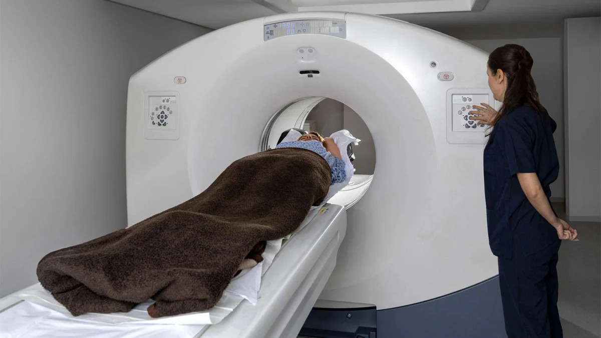
Throughout the trial, participants underwent both the novel 89Zr-DFO-daratumumab PET/CT and the standard-of-care 18F-FDG PET/CT. The safety profile was encouraging; researchers reported no severe adverse events associated with the tracer, with only minor, non-severe side effects noted in 5% of participants—primarily during the administration of the non-radiolabeled antibody component.
Supporting Data: A Tale of Two Scans
The trial results revealed a fascinating, nuanced landscape of diagnostic capability. When comparing the two modalities, the researchers found no statistically significant difference in overall disease detection: 43% for the CD38-targeted tracer versus 47% for the standard FDG scan (P=0.81).
However, looking at the aggregate data masks the most significant finding: the two scans are highly complementary. In a significant number of cases, the modalities identified lesions that the other completely missed:
- Unique Detection: 13% of patients had disease visualized only by the 89Zr-DFO-daratumumab tracer, while 17% of patients had disease visualized only by the standard FDG PET/CT.
- Superior Sensitivity: In 28% of the patient cohort, the CD38-targeted tracer identified a greater number of disease sites than the standard scan. Conversely, the standard FDG PET/CT was more sensitive in 22% of patients.
These data points underscore that neither technology is currently a "silver bullet," but together, they provide a much more comprehensive assessment of a patient’s systemic disease than either could achieve alone.
Official Perspectives and Expert Analysis
Dr. Ulaner emphasized the clinical utility of these findings in a discussion with MedPage Today. "This study demonstrates that immuno-PET targeting CD38 can identify, localize, and quantify myeloma, which is overlooked by FDG PET in many patients," he said. "This allows oncologists to optimally stage, target, and treat disease."
He acknowledged that the field of radiology is rarely characterized by perfection. "While no imaging modality is perfect, this adds a new tool for the care of patients with multiple myeloma," he added.
Co-author C. Ola Landgren, MD, PhD, of the Sylvester Comprehensive Cancer Center at the University of Miami, framed the discovery within the broader goal of a cure. "If we are going to cure multiple myeloma, we need two things. One, we need really effective medicines. Second, we need to be able to make sure there is no disease left behind after these treatments," Dr. Landgren noted in a press release. "We are developing new and better technology designed to see where the disease is and make sure we are not missing it. We show that you can successfully find much more disease."
In an editorial accompanying the publication, Peter Choyke, MD, of the National Cancer Institute, offered a balanced perspective. He noted that while it might be "disappointing" that neither agent could detect more than half of the disease in patients with histologically confirmed myeloma, there is profound reason for optimism. He highlighted the "theranostic" potential of the tracer: because the imaging agent is based on daratumumab, a positive scan provides a "companion diagnostic" function, suggesting that the patient may be a good candidate for daratumumab-based therapy.
Clinical Implications and Future Directions
The implications of this study are twofold. First, the integration of CD38-targeted imaging could fundamentally change how clinicians assess "minimal residual disease" (MRD). By using these tracers to verify that a treatment has truly cleared the body of myeloma, doctors can move beyond bone marrow biopsies, which only provide a small, localized snapshot of the disease.
Second, Dr. Choyke’s editorial suggests a future where molecular imaging is combined with liquid biopsies—blood-based tests that detect circulating tumor DNA. This "multi-modal" approach could potentially detect early disease recurrence months or even years before it would be visible on standard, structural imaging.
As the research community moves forward, the focus will likely shift to optimizing the timing and combination of these imaging techniques. The ability to "see" the disease with such specificity means that treatments can be tailored, escalated, or de-escalated based on real-time evidence of therapeutic response.
Ultimately, the development of 89Zr-DFO-daratumumab represents a significant leap forward in precision medicine. By providing a clearer, more targeted view of the myeloma landscape, this technology moves the needle closer to the ultimate clinical goal: a comprehensive, patient-specific strategy that ensures no malignant cell is left behind. As clinical trials continue, the medical community will be watching closely to see how this "new tool" integrates into the standard of care for one of the most challenging hematologic malignancies.