











For many individuals living with Ehlers-Danlos Syndrome (EDS), the diagnostic journey is punctuated by a frustrating paradox: the severity of their daily physical struggle is rarely reflected in the clinical reports they receive. A typical MRI report for an EDS patient often reads as follows: “Cervical spine: Unremarkable. Thoracic spine: Unremarkable. Lumbar spine: Slight bulging disc at L5-S1.”
These findings often feel disconnected from the reality of the patient’s experience—chronic pain, neurological deficits, and debilitating fatigue. When a patient’s symptoms are dismissed because their scans appear "normal," it creates a profound sense of medical gaslighting. However, the issue may not lie with the patient’s perception or the clinician’s interpretation, but rather with the fundamental physics of the diagnostic tool itself. Standard MRI technology, while a marvel of modern medicine, captures the human body in a horizontal, non-weight-bearing state. For a condition defined by connective tissue laxity and mechanical instability, this "at-rest" snapshot may be missing the most critical data points.
The Diagnostic Mismatch: Why Supine Imaging May Fail
The human spine is a complex, dynamic structure designed to support the body against the constant pull of gravity. In patients with Ehlers-Danlos Syndrome, the ligaments that stabilize the vertebral column are often compromised by faulty collagen, leading to joint hypermobility and structural instability.
When a patient lies down in a standard MRI scanner, the weight of the head and torso is removed from the spinal column. The joints "unload," and the spine is allowed to decompress. If a patient’s spinal instability is a dynamic, mechanical problem—triggered by the simple act of sitting at a desk or standing in a grocery line—the supine MRI will likely fail to capture the pathology. By imaging the patient in their least symptomatic state, clinicians are inadvertently creating a diagnostic mismatch. The patient is being evaluated for a functional, weight-bearing problem using a static, horizontal test.
Chronology of the Search for Dynamic Imaging
The medical community’s awareness of the limitations of supine imaging has evolved over the last several decades.
- Early 2000s: Clinicians specializing in connective tissue disorders began noting that patients with Chiari malformation and craniocervical instability (CCI) often reported symptom relief when lying down, suggesting that gravity played a significant role in their neurological symptoms.
- 2007: Seminal research by Milhorat et al. highlighted the syndrome of occipitoatlantoaxial hypermobility, underscoring how cranial settling could manifest only under specific gravitational stresses.
- 2015: Health Quality Ontario released an evidence-based analysis, marking one of the first formal institutional looks at the utility of positional MRI for EDS and related abnormalities.
- 2020–2025: As interest in CCI and EDS-related spinal issues grew, organizations like the CCI Foundation began advocating for the inclusion of dynamic imaging in diagnostic protocols.
- 2026: Recent systematic reviews, including those published in European Journal of Radiology Open, have begun to categorize the specific clinical utilities of upright MRI, providing a more robust framework for when this technology should be utilized.
Supporting Data and Technical Realities
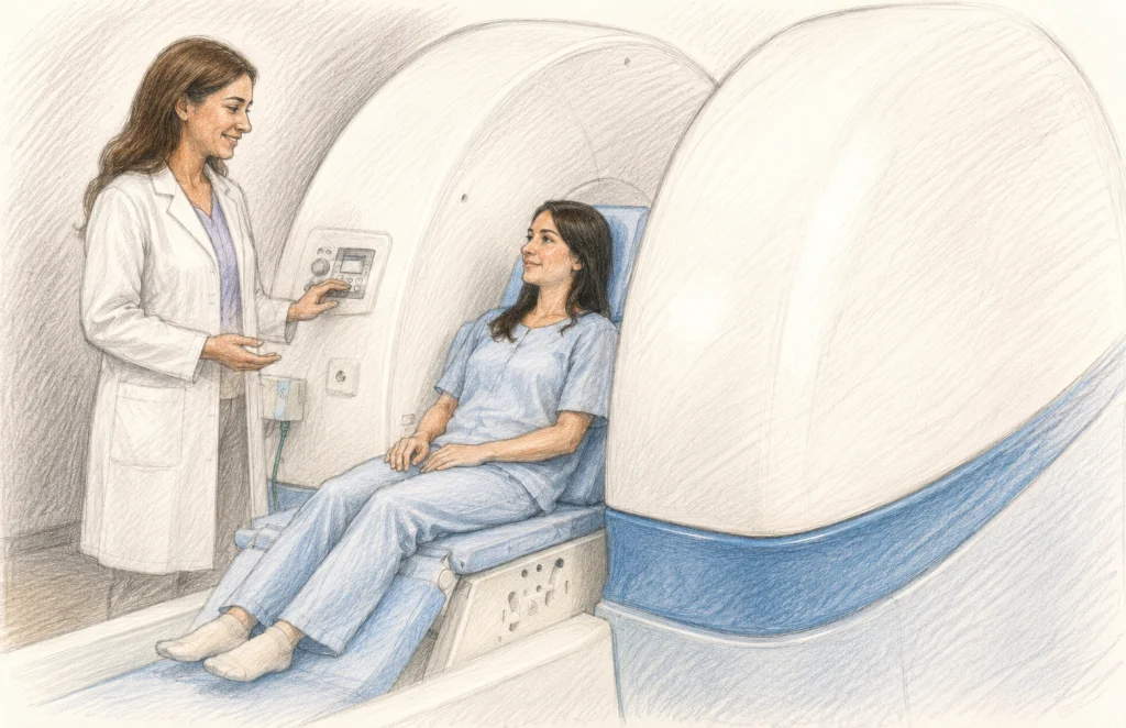
The push for upright, or positional, MRI (pMRI) is driven by the need to visualize the spine under the same mechanical loads that provoke pain. Upright systems allow patients to be imaged in various positions—flexion, extension, and rotation—while bearing their own weight.
The Trade-off of Field Strength
It is important for patients and clinicians to understand the technical constraints of current pMRI technology. Most standard hospital MRI scanners utilize high-field magnets (typically 1.5T or 3.0T), which provide exceptional image resolution. In contrast, most upright MRI systems currently operate at lower field strengths, usually between 0.6T and 1.0T.
This results in a trade-off: while the image quality may be slightly less granular than that of a high-field, closed-bore scanner, the diagnostic benefit of seeing the spine in its functional, weight-bearing state often outweighs the loss of image detail. For a surgeon deciding on a stabilization procedure, seeing a spinal cord being compressed during an upright posture is infinitely more valuable than a high-resolution, "normal" image of a relaxed, recumbent spine.
Evidence of Efficacy
A 2025 systematic review identified nine distinct studies confirming that dynamic and upright MRI could identify weight-bearing abnormalities that were entirely invisible in the supine position. These abnormalities included:
- Alterations in spinal alignment: Detecting subtle shifts in vertebrae that only occur under load.
- Narrowing of the spinal canal: Demonstrating how the spinal canal can stenose when the patient is upright.
- CSF Flow Dynamics: Assessing whether the flow of cerebrospinal fluid is obstructed at the craniocervical junction during head movement.
Official Perspectives and Clinical Recommendations
Medical societies are beginning to shift their stance on the necessity of dynamic imaging for complex spinal cases. While traditional imaging remains the "gold standard" for structural tumors or lesions, the clinical consensus for mechanical instability is shifting.
Organizations like the CCI Foundation now recommend that for patients whose symptoms are strongly influenced by posture (e.g., headaches that worsen upon standing, or limb weakness when looking upward), upright MRI should be considered a vital component of the diagnostic workup. However, it is not a replacement for traditional imaging. Rather, it acts as a functional "stress test" for the spine.
The Need for More Large-Scale Research
Despite the promising clinical utility of pMRI, experts caution that it is not a panacea. The medical community acknowledges a gap in large-scale, prospective studies that directly compare patient outcomes between those who receive pMRI and those who receive only standard imaging. Without these massive datasets, many insurance providers remain hesitant to cover the procedure, and many clinicians remain skeptical of its "added value."
Implications for Patients and Healthcare Providers
For the patient, the implications of this diagnostic gap are significant. The difficulty in obtaining an upright MRI often creates a barrier to care.
Access and Advocacy
Currently, access to pMRI is limited. These machines are not as ubiquitous as the scanners found in every major hospital emergency room. Patients often face:
- Geographic Barriers: The need to travel long distances to find a facility with upright capabilities.
- Financial Barriers: The high cost of out-of-pocket expenses if insurance deems the procedure "not medically necessary."
- Preauthorization Hurdles: The ongoing struggle to provide sufficient documentation to justify why a "lower field" scanner is required when a "high field" one is available locally.
For those navigating this, the best approach is a collaborative one. Patients should discuss with their physicians whether their specific symptom profile—specifically posture-dependent neurological changes—justifies the request for an upright scan.
When Should You Consider Upright MRI?
An upright MRI may be a relevant investigative tool if you experience:
- Symptoms that are consistently aggravated by standing, sitting, or prolonged upright posture.
- Neurological symptoms (numbness, tingling, weakness) that appear only during specific head or neck movements.
- A discrepancy where clinical symptoms suggest significant spinal pathology, but standard supine MRI reports remain "unremarkable."
- Suspected craniocervical instability or tethered cord syndrome where positional changes in the spine are expected.
Moving Toward a Holistic Diagnostic Model
The spine is not a static column; it is a complex, moving part of a biological machine. By viewing it only through the lens of static, supine imaging, we are observing only one part of a larger story. For those with Ehlers-Danlos Syndrome, the "normal" results on a traditional MRI are not a sign that nothing is wrong—they are a sign that we are looking at the problem from the wrong angle.
The integration of upright, dynamic MRI into standard practice represents a move toward more patient-centered, functional medicine. While the technology is currently hindered by issues of access and the need for more expansive research, the path forward is clear: to truly understand the spine in EDS, we must see it in motion. As awareness grows, the hope is that these diagnostic tools will become as accessible as they are essential, finally providing patients with the clarity they deserve.
Key Takeaways
- Positional Mismatch: EDS patients often suffer from mechanical instability that only manifests under the weight-bearing demands of daily life, making traditional "at-rest" MRIs potentially misleading.
- The Upright Advantage: Upright MRI allows for the assessment of the spine while the patient is sitting or standing, revealing potential compressions or misalignments that vanish when the patient lies down.
- Technical Compromise: Upright MRI machines generally offer lower image resolution (lower Tesla) than hospital-grade scanners, but they provide critical functional data that higher-resolution scans may miss.
- Advocacy and Access: Patients should work closely with specialists to navigate insurance and access issues, framing the scan as a "functional stress test" rather than a replacement for standard imaging.
- Future Direction: While the clinical utility is supported by growing evidence, more large-scale studies are required to establish upright MRI as a standard-of-care procedure across all medical institutions.