











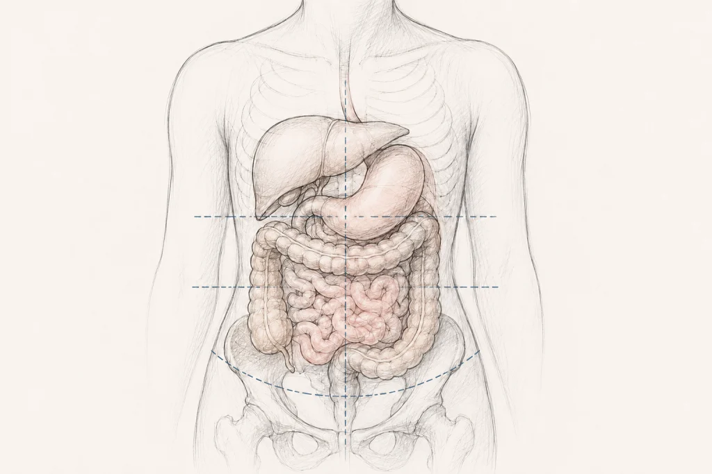
In the landscape of modern medicine, where diagnostic technology allows us to peer into the human body with unprecedented clarity, some conditions have managed to slip through the cracks of history. One such condition is visceroptosis—the downward displacement of internal organs from their normal anatomical positions. While pelvic organ and rectal prolapse are well-documented features of Hypermobile Ehlers-Danlos syndrome (hEDS), the descent of abdominal organs higher in the torso remains a medical "blind spot."
As we navigate an era of increased awareness regarding connective tissue disorders, the medical community is being forced to confront a decades-old question: Could the "forgotten" diagnosis of visceroptosis be the missing link in explaining the complex, multi-systemic gastrointestinal agony faced by thousands of patients today?
The Core Concept: What is Visceroptosis?
To understand visceroptosis, one must first look at the etymology of the term. Derived from the Latin viscera (internal organs) and the Greek ptosis (a falling or downward displacement), the term describes a state where organs—such as the stomach, liver, kidneys, or colon—migrate from their anatomically intended "homes" into the lower abdomen or pelvic cavity.
While the human body is designed with a degree of natural movement, visceroptosis implies a structural failure. In the context of connective tissue disorders, this is often attributed to the laxity of the ligaments and fascia that serve as the body’s internal "anchors." When these support structures lose their tensile strength, the organs they tether succumb to the relentless pull of gravity.
A Chronology of a Forgotten Diagnosis
The Era of "Glénard’s Disease" (1885–1930s)
In the late 19th century, French physician Dr. Frantz Glénard revolutionized the understanding of abdominal complaints. He observed a cohort of patients who suffered from chronic, debilitating abdominal distress that defied standard explanations of the day. He identified that these patients possessed organs sitting significantly lower in the abdominal cavity than what was considered standard anatomy.
For nearly 50 years, "Glénard’s disease" was a staple of clinical practice. It was not a fringe theory; it was a recognized medical diagnosis appearing in prestigious publications like The New England Journal of Medicine, JAMA, and The Lancet. Radiologists actively sought out organ displacement on early imaging, and surgeons attempted various corrective procedures.
The Great Disappearance
Sometime mid-20th century, the diagnosis fell out of clinical favor. The reasons remain largely absent from medical textbooks. Some historians of medicine suggest that as the field of psychiatry gained influence, the physical complaints of patients with visceroptosis were increasingly rebranded as "neurasthenia," "somatic dysfunction," or purely psychological anxiety. The focus shifted away from the physical displacement of organs toward the patient’s perceived emotional state.
The Modern Resurgence
In the 21st century, the conversation has reignited. As the Ehlers-Danlos syndrome (hEDS) community has grown, patients have consistently reported symptoms—severe bloating, gastroparesis-like nausea, and intractable constipation—that mirror the historical descriptions of Glénard’s patients. We are now in a period of re-discovery, where clinicians are beginning to ask if the "forgotten" diagnosis was simply ahead of its time.
Supporting Data: The Link to Connective Tissue Laxity
The primary barrier to acknowledging visceroptosis has been the lack of a standardized diagnostic protocol. However, recent literature provides a compelling foundation for further research.
Anatomic vs. Clinical Visceroptosis
A critical distinction must be made: Anatomic visceroptosis refers to the simple position of an organ, whereas clinical visceroptosis describes a symptomatic state where that displacement disrupts function. Many individuals may have "dropped" organs that remain asymptomatic. The clinical threshold is met only when the displacement directly contributes to physiological failure, such as obstruction, impaired motility, or chronic pain.
Findings in Modern Research
- The 2023 Study: A recent study involving 139 patients with slow-transit constipation identified a significant correlation between transverse colon ptosis and the severity of symptoms. While this study was not exclusive to the EDS population, it provides objective evidence that organ position impacts functional health.
- The hEDS Connection: The 2017 international classification for hEDS officially recognized pelvic organ and rectal prolapse. However, case reports continue to document gastroptosis (stomach descent) and multiple visceral ptoses in hEDS patients, suggesting that the current diagnostic criteria may only be capturing the "tip of the iceberg" regarding organ displacement.
Implications for the Patient Experience
The patient journey for those suffering from undiagnosed visceroptosis is often marked by gaslighting and clinical dismissal. Because symptoms like nausea, abdominal pain, and bloating are common to many functional gastrointestinal disorders, physicians often look for "standard" diseases—like Crohn’s, IBS, or Celiac—and stop searching when those tests return negative.
The "Somatic" Stigma
Historically, patients with visceroptosis were labeled "neurasthenic." Today, that has morphed into labels such as "somatic" or "psychosomatic." This pattern of dismissal is particularly prevalent among women with hEDS. When a patient reports that their abdomen feels "full" or "heavy," or that they feel their organs "dropping" when they stand, they are frequently told it is an anxiety manifestation. This institutional skepticism prevents the necessary imaging—often requiring upright or weight-bearing scans—that would reveal the structural reality of the displacement.
Clinical Challenges and Future Directions
The Diagnostic Vacuum
Why isn’t visceroptosis standardly diagnosed? The answer lies in the limitations of our current diagnostic architecture.
- Imaging Protocols: Most abdominal imaging (CT and MRI) is performed with the patient in a supine (lying flat) position. Gravity, however, is the primary driver of visceroptosis. If you image a patient while they are lying down, you may inadvertently "correct" the ptosis, masking the very issue causing the patient’s symptoms.
- Lack of Criteria: There is currently no unified, international set of criteria to define "how much" displacement is clinically significant.
The Treatment Frontier
Treatment remains a complex, high-stakes endeavor:
- Conservative Management: The first line of defense includes physical therapy aimed at core stabilization, the use of abdominal compression garments to provide artificial "support" for the abdominal wall, and dietary modifications to manage transit time.
- The Surgical Dilemma: Surgery for non-pelvic visceroptosis is controversial. While some patients report life-changing relief, the surgical community remains hesitant. In the context of hEDS, tissue fragility poses a significant risk for surgical complications, including poor wound healing and further destabilization of the abdominal wall. Consequently, surgery is often reserved as a last resort, following exhaustive conservative efforts.
Conclusion: A Call for Medical Re-Evaluation
The history of visceroptosis serves as a cautionary tale of how quickly a recognized condition can be erased from the collective medical consciousness. For patients living with the daily reality of connective tissue laxity, the "rediscovery" of this condition is not merely academic—it is a potential pathway to validation and relief.
Moving forward, the medical community must prioritize:
- Developing standardized, upright imaging protocols to accurately assess organ position.
- Expanding the diagnostic criteria for hEDS-related structural issues beyond the pelvic floor.
- Reframing the patient narrative to move away from the "somatic" stigma, treating abdominal complaints as valid physiological consequences of connective tissue failure until proven otherwise.
As we look toward the future, the integration of visceroptosis into the modern clinical lexicon is not just about updating a textbook—it is about restoring the dignity of care for patients who have spent decades waiting for medicine to look down and see what they have been describing all along.
Key Takeaways
- Definition: Visceroptosis is the downward displacement of internal organs caused by the failure of connective tissue support.
- Historical Context: Once a recognized medical diagnosis, it vanished from standard practice in the mid-20th century, leading to a long period of diagnostic neglect.
- The hEDS Connection: Increased clinical awareness of hEDS has brought the condition back into focus, as patients exhibit symptoms that align with historical records.
- Diagnostic Hurdles: Current imaging (supine position) often fails to capture the displacement, and there is a lack of standardized diagnostic criteria.
- Treatment: While conservative therapies are preferred, surgical options remain experimental and carry significant risks for those with underlying connective tissue fragility.