











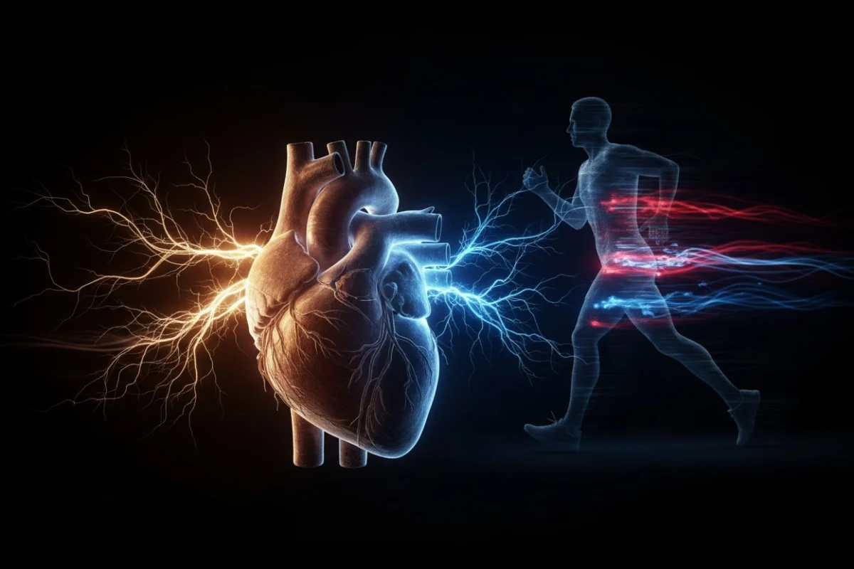
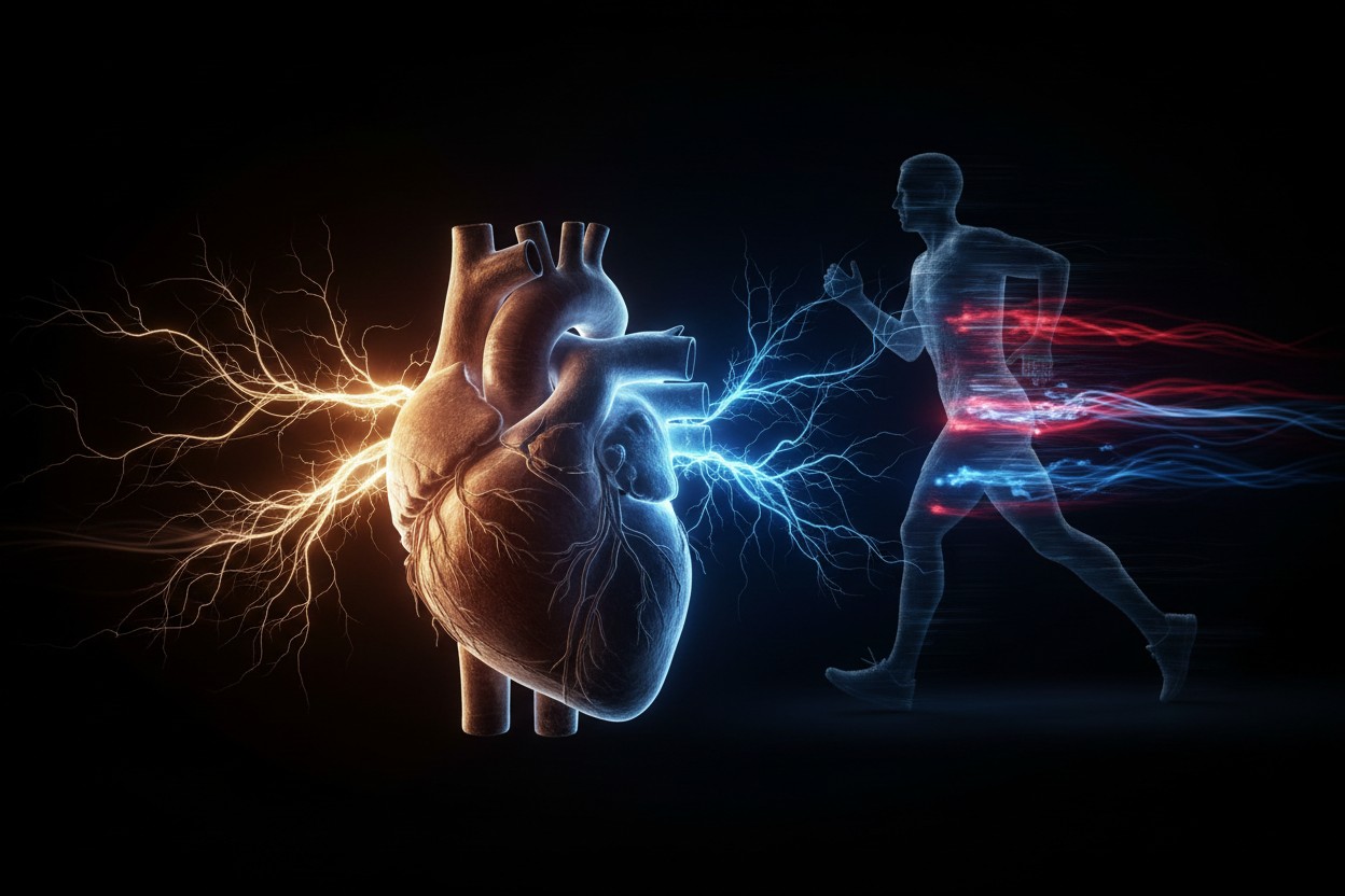
For decades, the medical community has operated under the mantra that exercise strengthens the heart muscle, improves circulation, and enhances endothelial health. However, a groundbreaking study published in the July 2026 issue of Autonomic Neuroscience suggests that the benefits of physical activity extend far deeper than the muscular walls of the heart. According to researchers at the University of Bristol, regular aerobic exercise physically reshapes the neural circuitry that acts as the heart’s "autopilot."
This discovery of asymmetric neuroplasticity in the stellate ganglia—the clusters of nerves responsible for transmitting "go faster" signals to the heart—challenges our fundamental understanding of how the autonomic nervous system adapts to physical stress. By revealing a distinct left-right pattern in how these nerves remodel themselves, the research opens a new frontier in the treatment of cardiovascular conditions ranging from chronic angina to stress-induced cardiomyopathy.
The Anatomy of the "Dimmer Switch"
To understand the significance of this research, one must first understand the role of the stellate ganglia. Located in the lower neck and upper chest, these paired nerve clusters are integral components of the sympathetic nervous system. They function as the body’s internal command center for cardiac performance, regulating heart rate and the force of contraction.
Dr. Augusto Coppi, a Senior Lecturer in Veterinary Anatomy at the University of Bristol and the lead author of the study, characterizes these nerve clusters as the "heart’s dimmer switch." While it has long been known that these nerves exert control over cardiac output, the Bristol team’s research is the first to demonstrate that exercise acts as a precise architect, remodeling this "switch" in a highly side-specific manner.
Chronology of the Discovery: A 10-Week Transformation
The study, which utilized advanced three-dimensional stereological imaging, followed a controlled group of rats through a 10-week aerobic training regimen. The methodology was designed to isolate the effects of repetitive cardiovascular demand on the autonomic nervous system.
Phase 1: Baseline Assessment
Before the exercise protocol commenced, researchers established a baseline for neural density and morphology in both the left and right stellate ganglia. At this stage, the neural architecture appeared relatively uniform, mirroring the standard anatomical models found in functional anatomy textbooks.
Phase 2: The Training Intervention
Over the course of 10 weeks, the subject rats engaged in a consistent, supervised aerobic training program. This phase was designed to simulate the physiological adaptations humans experience when transitioning from a sedentary lifestyle to a routine of regular cardiovascular exercise.
Phase 3: Post-Training Analysis
Upon completion of the 10-week period, the team utilized high-resolution 3D imaging to map the structural changes. The results were startling. The researchers observed significant, asymmetric remodeling:
- The Right Side: The right cardiovascular nerve cluster exhibited a four-fold increase in the number of neurons compared to untrained counterparts.
- The Left Side: While the right side prioritized quantity, the left side underwent a morphological shift. Neurons in the left stellate ganglia nearly doubled in size, while the overall cluster showed a subtle reduction in total neuron count.
This divergence suggests that the autonomic nervous system does not respond to exercise as a singular unit, but rather as a highly specialized, lateralized system.
Supporting Data: Why Asymmetry Matters
The findings from the University of Bristol provide the first empirical evidence of "left-right neuroplasticity" within the autonomic nervous system. This discovery is not merely academic; it addresses a long-standing curiosity in neuroanatomy regarding why the sympathetic supply to the heart is bifurcated.
In the context of cardiac physiology, the right and left stellate ganglia do not perform identical tasks. The right-sided nerves primarily influence the sinoatrial node, the heart’s natural pacemaker, while the left-sided nerves have a more pronounced impact on the ventricles and the heart’s contractile force. By disproportionately increasing the density of the right-sided nerves and the size of the left-sided neurons, the body appears to be "tuning" its neural hardware to optimize cardiac performance under stress.
This structural adaptation suggests that aerobic exercise induces a form of efficiency that has never been documented before. Instead of simply pushing the heart to work harder, the body is reconfiguring the communication lines to ensure that the heart responds more fluidly and resiliently to the demands of physical exertion.
Official Responses and Expert Perspective
The medical community has responded to the study with cautious optimism. Dr. Coppi, in his official statement, emphasized that while the findings are revolutionary, they represent the beginning of a long journey toward clinical application.
"We have uncovered a previously hidden pattern in the body’s autopilot system," Dr. Coppi stated. "By understanding these left-right differences, we aren’t just looking at anatomy; we are looking at a roadmap for personalized medicine."
Collaborators from University College London and the University of São Paulo echoed these sentiments, noting that the study bridges the gap between basic neuroanatomy and clinical cardiology. The inclusion of international partners highlights the global significance of the research, as researchers look to validate these findings across different animal models before moving toward human trials.
Implications for Modern Medicine
The clinical implications of this "neural rewiring" are vast, particularly for conditions that have historically been difficult to manage.
1. Arrhythmia Management
Cardiac arrhythmias are often caused by electrical instability in the heart, frequently triggered by erratic sympathetic signals. If doctors can understand how exercise influences the remodeling of the stellate ganglia, they may be able to develop non-invasive "neural training" protocols to stabilize heart rhythms in patients prone to tachycardia or atrial fibrillation.
2. Targeted Nerve Blocks
Currently, procedures such as stellate ganglion blocks are used to treat chronic pain and certain heart rhythm disorders. However, these procedures are often non-specific. The Bristol study suggests that because the left and right nerves serve different functions, a "one-size-fits-all" approach to denervation or blockade may be suboptimal. Identifying which side is contributing to a patient’s pathology could allow for more precise, effective interventions.
3. Stress-Induced Cardiomyopathy
"Broken-heart" syndrome, or Takotsubo cardiomyopathy, is a temporary heart condition brought on by extreme emotional stress, which causes a surge of catecholamines that "stuns" the heart. By understanding how the sympathetic chain adapts to training, researchers hope to identify how to "train" the nerves to better manage stress-induced signals, potentially protecting the heart from the damage caused by acute emotional trauma.
Limitations and the Path Forward
Despite the enthusiasm surrounding these findings, the research team is adamant about the need for further investigation. The study was conducted in rats, and the translation from rodent models to human clinical practice is complex.
"We must be careful not to overstate the current reach of this data," the report notes. "The neural complexity of the human autonomic system is significantly higher than that of our animal models."
The next steps for the research team include:
- Human Biomarkers: Developing non-invasive markers to observe these structural changes in human patients who undergo long-term exercise programs.
- Functional Correlation: Testing whether the structural changes in the stellate ganglia correlate with measurable improvements in heart rate variability (HRV) and recovery time after intense exercise.
- Cross-Species Validation: Investigating whether the same left-right asymmetry appears in larger mammals, which would provide a more accurate proxy for human physiology.
Conclusion: A New Foundation for Cardiology
The study from the University of Bristol represents a fundamental shift in how we view the relationship between exercise and the brain-heart axis. For years, we have treated the heart as a muscle that responds to stress; we must now begin to view it as a sophisticated biological engine whose "on-board computer" is constantly being reprogrammed by the activity we demand of it.
As we move toward a future of personalized medicine, the ability to map and potentially influence the stellate ganglia could provide doctors with a powerful new tool in their arsenal. By moving beyond traditional pharmaceuticals and surgical interventions, and toward therapies that leverage the body’s inherent ability to rewire its own neural architecture, the medical field may be on the cusp of a new era of cardiovascular health.
The research serves as a potent reminder that the human body is not a static machine, but a dynamic, learning system. Every step taken, every cycle completed, and every aerobic session logged is, quite literally, leaving a mark on the nervous system—a silent, asymmetric masterpiece of biological adaptation that keeps the heart beating in rhythm with the life we lead.