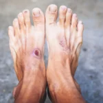
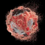





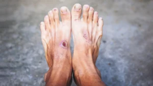
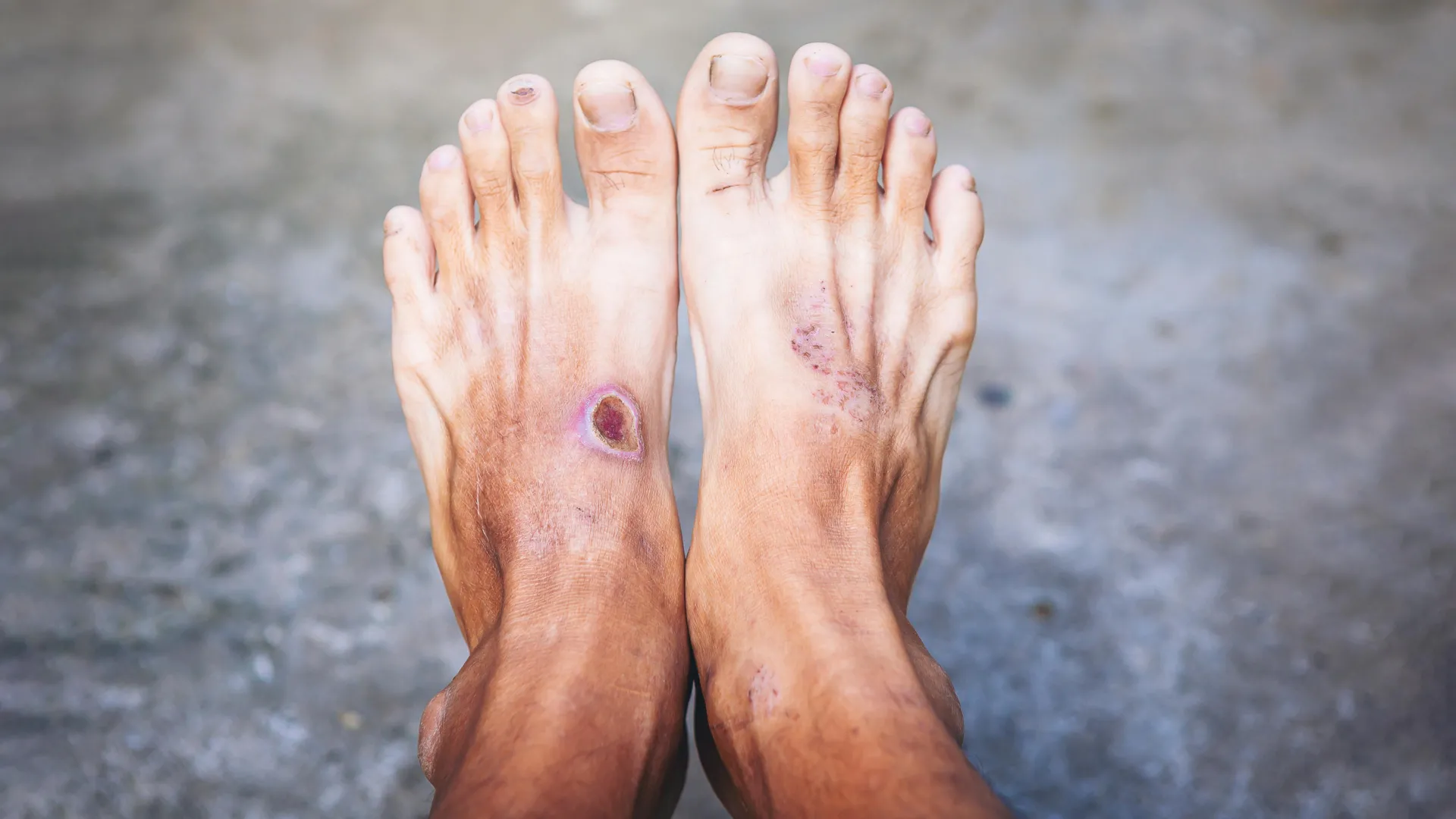
Chronic diabetic ulcers represent one of the most significant and costly challenges in modern medicine. Affecting over 131 million people globally, these wounds are not merely surface-level injuries; they are systemic failures of the body’s repair machinery. With associated healthcare costs reaching an estimated $755 billion annually, the burden of diabetic foot ulcers and similar complications is immense. Beyond the financial strain, these wounds carry the constant, looming threat of amputation and systemic infection, often leading to mortality.
Why do these wounds refuse to heal? A comprehensive new review led by Yi Ru and colleagues suggests the answer lies in the complex, often dysfunctional, immune landscape of the diabetic patient. By mapping the behavior of immune cell populations throughout the wound-healing trajectory, researchers are beginning to understand how diabetes "reprograms" the immune system, turning a life-saving defense mechanism into a barrier to recovery.
The Immune Landscape: A Chronology of Dysfunction
The process of wound healing is a tightly orchestrated symphony of biological events, typically categorized into three phases: inflammation, proliferation, and remodeling. In a healthy individual, the immune system acts as the conductor. However, in the diabetic wound, this symphony is derailed, resulting in a state of chronic, unresolved inflammation.
The Initial Phase: Neutrophils and Mast Cells
Immediately following an injury, the body rushes neutrophils to the site. Their primary mission is to eradicate pathogens and clear cellular debris. In a healthy wound, this is a transient event. In a diabetic wound, however, this phase becomes a trap. Research indicates that the formation of neutrophil extracellular traps (NETs)—web-like structures used to catch bacteria—is poorly regulated in diabetic patients. This leads to the excessive release of proteases and reactive oxygen species, which, instead of clearing infection, cause secondary collateral damage to the surrounding healthy tissue, effectively sustaining the inflammatory fire.
Simultaneously, mast cells—which release histamine and various growth factors—exhibit abnormal degranulation. In the diabetic environment, this hyper-activation contributes to prolonged inflammation, disrupting blood vessel permeability and preventing the transition into the next stage of healing.
The Mid-Phase: The Macrophage Stalemate
Perhaps the most critical "check-point" in healing is the transition of macrophages from the pro-inflammatory M1 state to the pro-reparative M2 state. Under normal physiological conditions, monocytes arrive at the wound and differentiate into M1 macrophages to fight infection, then seamlessly pivot to an M2 phenotype to initiate tissue repair and remodeling.
In diabetic wounds, this pivot fails. Macrophages become "locked" in the M1 state. This chronic pro-inflammatory profile prevents the wound from ever entering the proliferative phase. The tissue remains trapped in a state of high oxidative stress and inflammatory signaling, rendering the wound stagnant and resistant to traditional topical treatments.
The Late-Phase: Dendritic and T-Cell Impairment
As the wound progresses, dendritic cells and T cells are expected to manage the transition toward tissue closure. Dendritic cells are responsible for clearing apoptotic (dead) cells through a process called efferocytosis. Evidence suggests that in diabetic patients, this process is significantly impaired, partly due to the reduced activity of the SLC7A11 transporter. When dead cellular material is not cleared, it continues to act as an "alarm," signaling the immune system that the wound is still under attack, further reinforcing the inflammatory cycle.
Meanwhile, specialized T cells—such as regulatory T cells (Tregs) and dendritic epidermal T cells—show a marked decline in function. Tregs are essential for suppressing excessive immune activity and signaling for tissue remodeling. Their absence, combined with the reduced production of growth factors like insulin-like growth factor 1 (IGF-1) by epidermal T cells, means the structural components of the skin (keratinocytes) fail to multiply and close the wound.
Supporting Data: Emerging Players and Cellular Dynamics
While macrophages and neutrophils have long been the focus of wound research, newer data is highlighting the influential roles of B cells and natural killer (NK) cells.
Recent studies indicate that B cells are not merely bystanders; they actively influence the healing environment by modulating macrophage polarization. When recruited correctly, B cells can help shift the macrophage population toward the reparative M2 state, potentially breaking the inflammatory stalemate. Similarly, NK cells, through their interactions with other immune cells and their production of cytokines, appear to play a vital role in regulating the formation of new blood vessels (angiogenesis).
The cumulative data suggest that the failure to heal is not due to a single rogue cell type, but rather a cascading breakdown in the communication between various immune populations. The inability of these cells to "hand off" their responsibilities at the right time is the fundamental pathology of the diabetic ulcer.
Official Perspectives: Shifting the Treatment Paradigm
The medical community is increasingly viewing diabetic ulcers as an "immune-regulatory disorder" rather than a localized skin problem. This shift in perspective is driving a wave of innovation in clinical research.
Experts emphasize that the future of wound care lies in "reprogramming" the immune response. Several strategies are currently undergoing rigorous scrutiny:
- Topical Biologics: New therapies designed to intercept persistent inflammatory signaling are being developed to "mute" the excessive cytokine production that keeps wounds in an M1-locked state.
- Cell-Based Therapies: Mesenchymal stem cell (MSC) therapies and extracellular vesicles derived from these cells are gaining traction for their broad, systemic ability to restore immune homeostasis.
- Advanced Biomaterials: "Smart dressings" are being engineered to act as drug-delivery systems, releasing immune-modifying agents in response to the specific chemical cues of the wound bed.
- Precision Engineering: Technologies like Janus liposomes, which regulate redox balance, and IL-15 superagonists, which specifically boost the activity of dendritic epidermal T cells, represent the cutting edge of regenerative medicine.
Implications for Future Clinical Practice
The most significant takeaway from the research by Yi Ru and colleagues is the critical importance of timing. A therapy that is highly effective during the initial inflammatory phase might be entirely ineffective—or even detrimental—if applied during the remodeling phase.
Personalized Immune Profiling
The implications for future healthcare are clear: "one-size-fits-all" wound care is becoming obsolete. The future of treating diabetic ulcers will likely involve personalized immune profiling. By taking a sample of the wound fluid and analyzing the dominant immune cell populations, clinicians could determine the exact biological stage of the wound and select the corresponding treatment.
Combination Therapies
Because the dysfunction involves multiple cell types, researchers are beginning to favor combination therapies. A dual-action approach—for example, using a drug to force M1-to-M2 macrophage conversion while simultaneously using an agonist to stimulate T-cell-mediated growth factor production—may provide the best chance for closure in recalcitrant wounds.
Addressing the Knowledge Gap
Despite these advancements, questions remain. The scientific community continues to debate the rigidness of the M1/M2 macrophage classification, with some arguing for a more nuanced, "spectrum-based" model of immune behavior. Resolving these debates is not just an academic exercise; it is essential for refining the next generation of drugs.
Conclusion
Chronic diabetic ulcers remain a formidable adversary, but our understanding of the battlefield is rapidly evolving. We are moving away from seeing these wounds as "lazy" or "neglected" injuries and toward understanding them as complex, immunological failures. By decoding how immune cells interact, fail, and potentially recover, science is laying the groundwork for a new era of wound care.
The path forward requires a transition toward precision medicine, where the treatment is as dynamic as the wound itself. As we unlock the secrets of immune signaling, we move closer to providing patients with a definitive solution for these life-altering complications, potentially saving millions from the cycle of chronic pain, disability, and systemic illness. The integration of immunological insights into clinical practice is not just a scientific ambition; it is an urgent public health necessity.