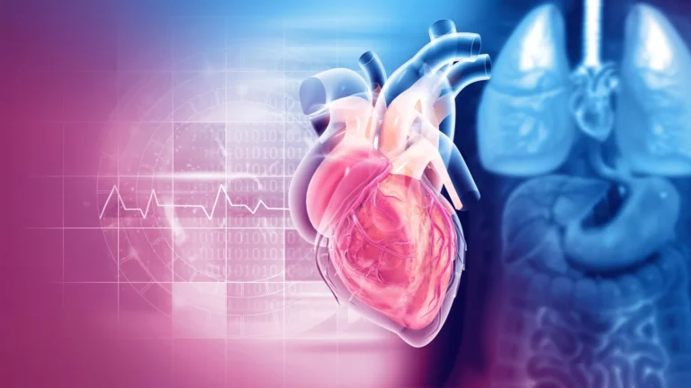











In a medical landscape where chronic stress has long been considered an intangible, subjective experience, researchers have achieved a breakthrough that promises to bring the silent toll of stress into the clinical spotlight. A team from the Johns Hopkins University School of Medicine has successfully utilized a deep learning artificial intelligence model to identify the first biomarker of chronic stress visible on standard medical imaging.
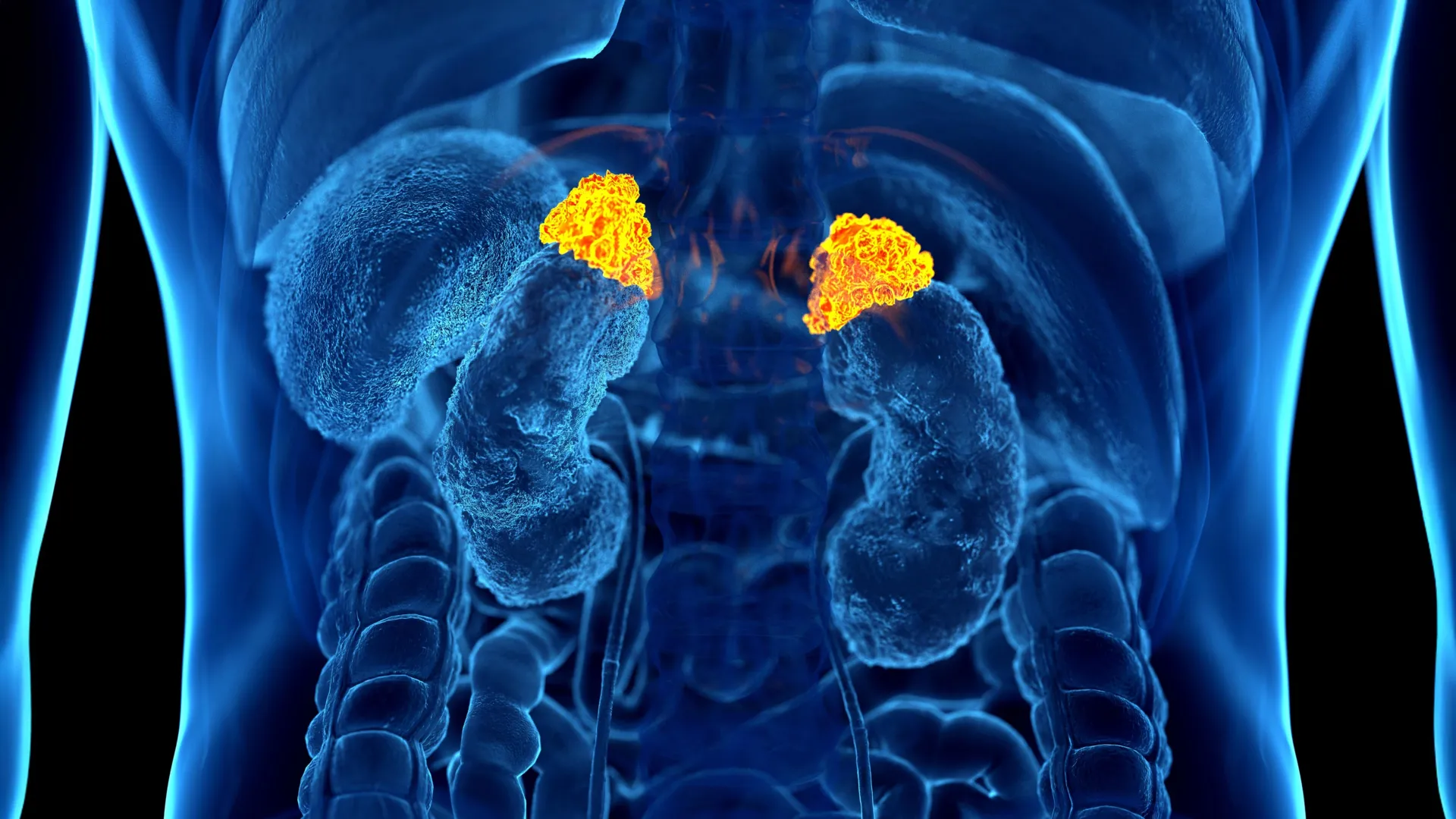
By analyzing routine chest CT scans, the AI can measure the volume of the adrenal glands—the organs responsible for the body’s "fight or flight" response—to quantify the cumulative, long-term impact of stress on the human body. This discovery, set to be unveiled at the upcoming annual meeting of the Radiological Society of North America (RSNA), marks a paradigm shift in how clinicians might soon monitor, diagnose, and treat the systemic health consequences of modern life.
Main Facts: Measuring the "Physiological Cascade"
Chronic stress is far more than a psychological state; it is a systemic physiological phenomenon. According to the American Psychological Association, the condition is a known contributor to anxiety, insomnia, muscular pain, hypertension, and immune system suppression. More severely, it has been clinically linked to heart disease, clinical depression, and obesity.
Until now, the "allostatic load"—the wear and tear on the body caused by chronic stress—has been notoriously difficult to measure. Previous methods relied on subjective patient questionnaires or cumbersome laboratory tests, such as measuring cortisol levels in serum or saliva. These tests often provide only a snapshot of a patient’s stress levels at a single moment in time.
The new AI-driven approach, led by Dr. Elena Ghotbi, M.D., a postdoctoral research fellow at Johns Hopkins, changes this by utilizing the Adrenal Volume Index (AVI). The researchers theorize that because the adrenal glands enlarge in response to prolonged stress, they act as a "biological gauge" of long-term physiological pressure. By applying a deep learning algorithm to existing chest CT scans—millions of which are performed annually in the United States—doctors can now extract this data without the need for additional radiation or invasive diagnostic procedures.
Chronology: From Data Collection to Clinical Validation
The development of this biomarker was the result of a rigorous, multi-year scientific investigation. The researchers utilized data from 2,842 participants (mean age 69.3; 51% women) who were part of the Multi-Ethnic Study of Atherosclerosis (MESA).
The stages of this research included:
- Cohort Integration: The study relied on the MESA cohort, which is uniquely comprehensive. It combined chest CT imaging, validated stress questionnaires, and rigorous physiological data, including glucose levels, white blood cell counts, heart rate, blood pressure, and albumin levels.
- Algorithmic Training: The research team trained a deep learning model to automatically outline and segment the adrenal glands from the CT scans.
- Developing the Index: The team established the Adrenal Volume Index (AVI) as a normalized metric: adrenal volume (cm³) divided by height squared (m²).
- Correlation Testing: Researchers compared the AVI against salivary cortisol levels—collected eight times a day over two days—and against psychosocial stress indicators, such as depression scores.
- Longitudinal Follow-up: The team tracked participants for up to a decade, correlating their AI-derived AVI with clinical outcomes such as heart failure and mortality.
Supporting Data: The Link Between Adrenal Size and Heart Health
The statistical findings of the study provide compelling evidence that adrenal gland enlargement is a direct proxy for chronic stress and, consequently, cardiovascular risk.
The study found that higher AVI values correlated significantly with:
- Hormonal Exposure: Higher overall cortisol exposure and higher peak cortisol levels.
- Psychological Indicators: Individuals reporting high perceived stress consistently displayed higher AVI than those who reported low stress.
- Structural Cardiovascular Changes: Increased AVI was linked to a higher left ventricular mass index, a key indicator of heart structure changes that often precede heart failure.
Perhaps most critically, the researchers discovered a dose-dependent relationship between the index and adverse outcomes. For every 1 cm³/m² increase in AVI, the risk of heart failure and death increased proportionately. This confirms that the AI-derived metric is not merely a theoretical construct, but a validated clinical predictor of major health events.
Official Responses and Expert Commentary
The research team emphasizes that this discovery is not intended to replace existing medical diagnostics, but rather to augment them with a practical, low-cost tool.
"Our approach leverages widely available imaging data and opens the door to large-scale evaluations of the biological impact of chronic stress," said Dr. Ghotbi. "This AI-driven biomarker has the potential to enhance cardiovascular risk stratification and guide preventive care without additional testing or radiation."
Dr. Shadpour Demehri, a professor of radiology at Johns Hopkins and the senior author of the study, highlighted the ease with which this could be integrated into existing hospital workflows. "For the first time, we can ‘see’ the long-term burden of stress inside the body, using a scan that patients already get every day," Dr. Demehri stated. "Until now, we haven’t had a way to measure and quantify the cumulative effects of chronic stress, other than questionnaires or serum markers, which are very cumbersome to obtain."
Teresa E. Seeman, Ph.D., a professor of epidemiology at UCLA and a pioneering researcher in the field of stress and health, provided an external perspective on the study’s impact. "What makes this work so exciting is that it links a routinely obtained imaging feature—adrenal volume—with validated biological and psychological measures of stress," Dr. Seeman noted. "It is a true step forward in operationalizing the cumulative impact of stress on health."
Implications: A New Frontier in Preventive Medicine
The implications of this breakthrough are vast, particularly for aging populations. By identifying "stress-burdened" patients through routine screenings, clinicians may be able to intervene earlier, prescribing lifestyle modifications, therapy, or pharmaceutical management before chronic stress manifests as irreversible cardiovascular damage.
Furthermore, the study provides a robust validation of the concept of "allostatic load." By moving stress from the category of "subjective mood" into the category of "measurable biological data," the research empowers doctors to treat the root causes of systemic inflammation and heart disease more effectively.
The research team, which includes co-authors from institutions like UCLA and the National Institutes of Health, believes this tool could be applied to a variety of stress-related diseases. As the healthcare industry continues to embrace the potential of artificial intelligence, this "imaging biomarker" stands as a prime example of how machine learning can transform existing medical data into actionable, life-saving insights.
As the medical community convenes at the RSNA, the conversation around this technology will likely focus on how quickly it can be standardized across radiology departments globally. With the infrastructure already in place—the millions of chest CTs already being performed—the transition from research to clinical standard could be faster than any previous diagnostic innovation in the field of stress management.