











For decades, the medical community has viewed chronic stress as a silent, elusive adversary. While patients often describe the sensation of being “stressed out,” quantifying that burden has historically relied on subjective questionnaires or cumbersome hormonal testing. However, a groundbreaking study set to be presented at the upcoming Radiological Society of North America (RSNA) annual meeting promises to change that, as researchers have successfully utilized deep learning artificial intelligence to identify the first-ever biomarker of chronic stress visible on standard medical imaging.
By leveraging routine chest CT scans—millions of which are performed annually across the United States—researchers at the Johns Hopkins University School of Medicine have developed a way to visualize the long-term, cumulative physiological toll of stress on the human body.
The Science of the "Stress Gauge"
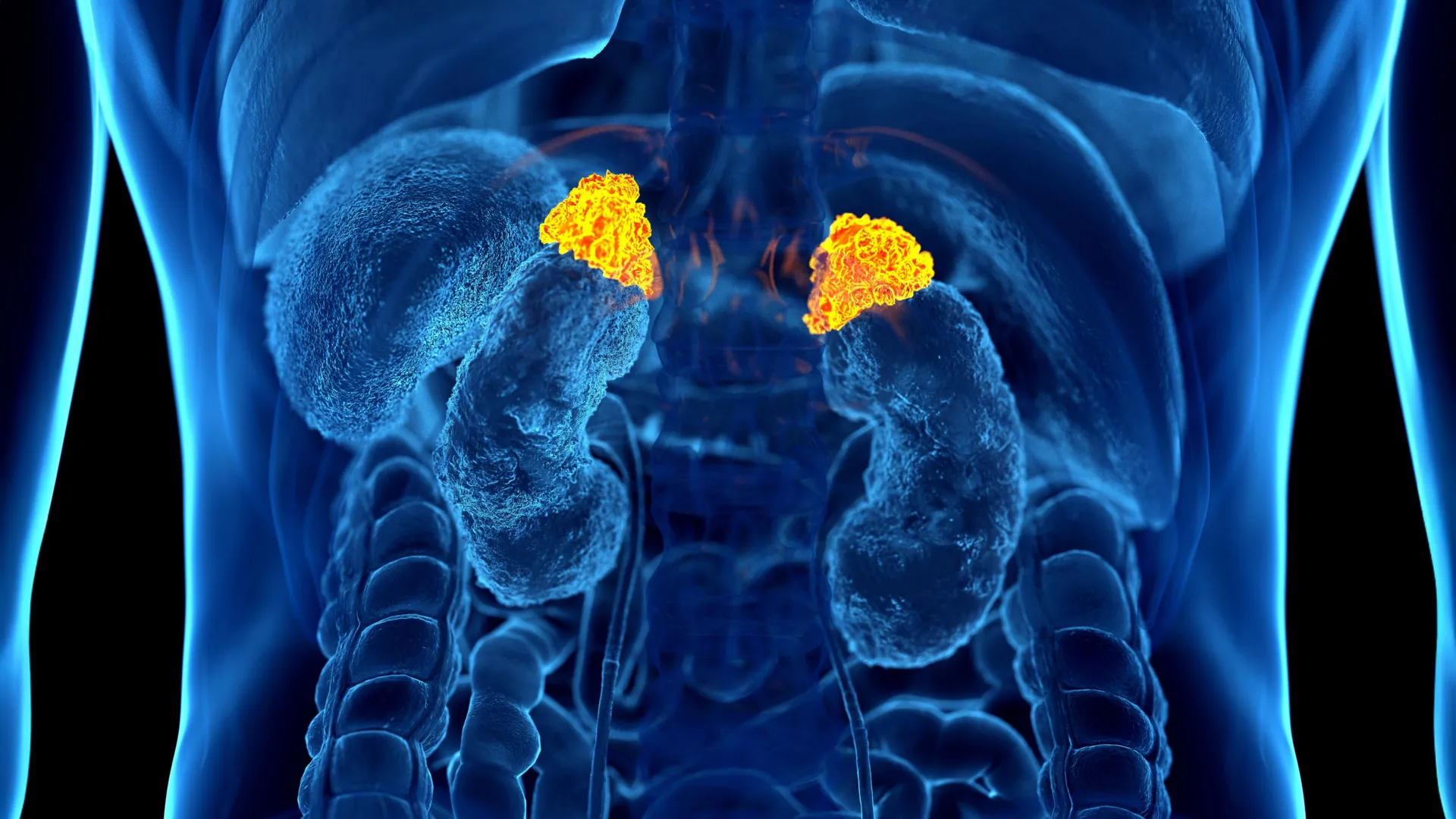
The core of this breakthrough lies in the adrenal glands, the small, triangular organs located on top of the kidneys. These glands are the primary command centers for the body’s stress response, releasing hormones like cortisol that govern our "fight or flight" mechanism.
While a standard blood or saliva test for cortisol can provide a snapshot of stress at a single, fleeting moment in time, the adrenal glands themselves act as a more permanent record. Much like a muscle that grows with exercise, the adrenal glands can undergo structural changes in response to the chronic, relentless activation of the body’s stress pathways.
Dr. Elena Ghotbi, M.D., a postdoctoral research fellow at Johns Hopkins and lead author of the study, spearheaded the development of a deep learning model capable of automatically segmenting and calculating the volume of these glands on existing CT imagery. By defining an “Adrenal Volume Index” (AVI)—a ratio of adrenal volume to a patient’s height—the team has created a stable, measurable indicator of long-term physiological stress.
Chronology of a Breakthrough
The journey to this discovery was rooted in a massive, multi-year epidemiological effort. The research team analyzed data from 2,842 participants, with a mean age of 69.3, drawn from the Multi-Ethnic Study of Atherosclerosis (MESA).
- Cohort Selection: The team leveraged the MESA study precisely because it was one of the few longitudinal datasets in the world that integrated three distinct pillars of health data: standardized chest CT imaging, validated psychosocial stress questionnaires, and comprehensive biochemical measurements, including salivary cortisol and indicators of "allostatic load."
- AI Model Training: The investigators applied their deep learning architecture to the CT scans of these participants. The AI was tasked with outlining the adrenal glands and automatically calculating the AVI, effectively turning thousands of raw, static images into actionable data points.
- Biological Correlation: Once the AVI was established for each patient, the team compared these values against the patients’ historical records—including their cortisol profiles, blood chemistry (glucose, creatinine, hemoglobin, etc.), and reported psychosocial stress levels.
- Outcome Validation: Finally, the researchers mapped these AVI values against long-term clinical outcomes. With up to 10 years of follow-up data available for the participants, the team could correlate the AI-derived stress marker with actual incidences of heart failure and cardiovascular mortality.
Supporting Data: Connecting the Dots
The findings revealed a robust statistical link between adrenal volume and the physical consequences of chronic stress. The data demonstrated that higher AVI values were consistently associated with:
- Elevated Hormonal Exposure: Patients with higher AVI had greater overall cortisol exposure and higher peak cortisol levels throughout the day.
- Increased Allostatic Load: Allostatic load—the "wear and tear" on the body resulting from chronic over-activation of physiological systems—was markedly higher in those with larger adrenal volumes.
- Psychosocial Alignment: The AI-measured AVI values aligned closely with established subjective stress indicators, such as patient-reported depression scores and perceived stress surveys.
- Cardiovascular Risk: Perhaps most critically, the study found that for every 1 cm³/m² increase in AVI, the risk of heart failure and death rose significantly. Furthermore, higher AVI was tied to an increased left ventricular mass index, a key indicator of adverse changes in heart structure.
Official Perspectives from the Research Frontline
The implications of this study are being met with significant enthusiasm within the medical community. Dr. Shadpour Demehri, M.D., professor of radiology at Johns Hopkins and senior author of the study, emphasized the clinical utility of the discovery.
“For the first time, we can ‘see’ the long-term burden of stress inside the body, using a scan that patients already get every day in hospitals across the country,” Dr. Demehri noted. “Until now, we haven’t had a way to measure and quantify the cumulative effects of chronic stress, other than questionnaires or surrogate markers that are often cumbersome to obtain.”
Dr. Teresa E. Seeman, Ph.D., a professor of epidemiology at UCLA and a pioneering researcher in the field of stress and health, co-authored the study and underscored the transformative nature of the work. “For over three decades, we’ve known that chronic stress can wear down the body across multiple systems,” she stated. “What makes this work so exciting is that it links a routinely obtained imaging feature—adrenal volume—with validated biological and psychological measures of stress. It is a true step forward in operationalizing the cumulative impact of stress on health.”
Implications for Future Clinical Practice
The potential impact of this AI-driven biomarker on public health is immense. Because chest CT scans are among the most common diagnostic tools in modern medicine, the infrastructure for this assessment already exists.
Enhancing Preventive Care
Rather than requiring patients to undergo new, expensive, or invasive testing, the AVI could be generated as a "bonus" metric during standard diagnostic procedures. If a patient undergoes a chest CT for a lung concern, the AI could simultaneously provide a "stress score," allowing clinicians to initiate preventive cardiovascular care or mental health interventions earlier than current protocols allow.
Cardiovascular Risk Stratification
The ability to link an imaging marker to independent clinical outcomes like heart failure offers a new layer of risk stratification. It allows doctors to see beyond cholesterol levels and blood pressure, capturing the hidden, biological impact of the patient’s lived experience and environment.
A Scalable Tool for Chronic Disease
As middle-aged and older populations face an increasing burden of stress-related ailments—ranging from hypertension and obesity to depression and immune dysfunction—the AVI offers a scalable, objective, and physiologically grounded method for tracking disease progression. By identifying patients with high "stress loads" before they experience a major cardiac event, the medical community may finally be able to transition from reactive treatment to proactive, personalized care.
The research team, which includes a multidisciplinary group of experts such as Dr. Joao A.C. Lima and Dr. Matthew J. Budoff, represents a significant collaborative effort to bridge the gap between radiology, endocrinology, and cardiovascular medicine. As the RSNA meeting approaches, the medical community is poised to take a definitive step toward making the "silent killer" of chronic stress visible—and, ultimately, manageable—in the modern clinical landscape.