










For decades, modern medicine and public health discourse have framed body fat as a biological adversary—a surplus to be shed in the pursuit of longevity and disease prevention. However, a groundbreaking study published by a multi-disciplinary team at the University of Michigan has fundamentally shifted this narrative. By investigating the rare, devastating condition known as familial partial lipodystrophy type 2 (FPLD2), researchers have confirmed that adipose tissue is not merely a storage depot for excess energy, but a sophisticated, active organ essential for systemic metabolic homeostasis.
The findings, led by a collaborative team of clinicians and basic science physiologists, suggest that when fat cells fail, the resulting metabolic collapse mimics, and in some cases exceeds, the damage caused by obesity. This discovery forces a re-evaluation of how we categorize metabolic disorders, moving the spotlight from the pancreas toward the complex, often-misunderstood world of the adipocyte.
The Biological Paradox: Why Fat is More Than Energy Storage
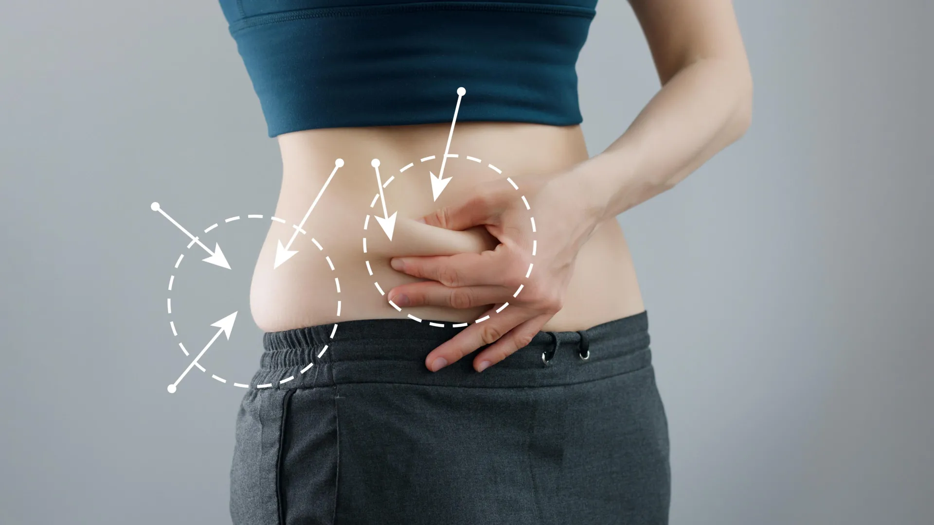
To the average observer, fat tissue (adipose tissue) is static—a reservoir of stored calories. In reality, it is a dynamic endocrine organ that secretes hormones, regulates systemic inflammation, and serves as a critical buffer for lipid distribution.
The Dual Risks of Metabolic Extremes
The medical community has long understood that excessive adiposity is a primary driver of Type 2 diabetes, cardiovascular disease, and chronic inflammation. Yet, the inverse—the pathological loss of fat—presents a metabolic catastrophe that is equally, if not more, severe.
In patients with FPLD2, a rare genetic disorder caused by mutations in the lamin A/C gene, the body loses the ability to maintain healthy fat deposits. As this tissue vanishes or redistributes abnormally, patients develop severe metabolic complications, including insulin resistance, hypertriglyceridemia, and fatty liver disease. Elif Oral, M.D., a professor in the Division of Metabolism, Endocrinology and Diabetes, has spent a significant portion of her career grappling with this paradox: if fat is the "bad guy," why does its absence trigger such profound metabolic disease?
Chronology of the Investigation
The quest to resolve this mystery required a bridge between the clinic and the laboratory.
Phase I: Clinical Observation
Dr. Oral began by observing the clinical trajectory of patients suffering from FPLD2. These patients often presented with a paradoxical profile: despite having very low levels of body fat, they exhibited the metabolic markers of advanced obesity-related diabetes. It became clear that the metabolic dysfunction was not a consequence of having too much fat, but rather a consequence of the failure of fat tissue to function as an endocrine organ.
Phase II: The Molecular Bridge
To understand the underlying mechanisms, Dr. Oral partnered with Ormond MacDougald, Ph.D., a professor of Molecular & Integrative Physiology. They were joined by a large, collaborative team, including graduate student researcher Jessica Maung, Ph.D. The team sought to replicate the clinical phenotype in a controlled setting.
They developed a sophisticated mouse model specifically engineered to "switch off" the lamin A/C gene within adipocytes. This allowed the researchers to observe the step-by-step decay of fat cells, providing a window into the "catastrophic" internal environment of cells that lose their structural integrity.
Phase III: Comparative Analysis
The researchers took the process a step further by comparing their mouse model data against actual tissue donated by human patients. The alignment between the animal model and human physiology provided the researchers with a high-resolution view of the disease’s progression, identifying the precise moment that healthy, functional fat begins its transition into a diseased state.
Supporting Data: When Cells Stop Functioning
The data gathered from the study revealed a "perfect storm" of cellular dysfunction that eventually leads to the total loss of adipose tissue.
The Breakdown of Lipid Metabolism
The researchers identified major disruptions in gene activity. Normally, adipocytes act as a high-capacity warehouse, taking in lipids and storing them safely. In the diseased cells, this process failed entirely. Unable to process or store lipids, the cells became bloated and toxic, ultimately leading to their destruction.
Mitochondrial Failure and Pro-Inflammatory Shifts
The study provided critical insights into the internal health of the cell:
- Mitochondrial Decay: Mitochondria, the powerhouses of the cell, failed to generate energy effectively. This loss of cellular respiration crippled the adipocyte’s ability to maintain its basic functions.
- The Inflammatory State: Both the adipocytes and the surrounding immune cells shifted into a state of chronic, pro-inflammatory activation. This inflammation acts as a toxic feedback loop, signaling neighboring cells to degrade and creating a systemic environment that promotes insulin resistance.
As Dr. Maung noted, these effects do not act in isolation. "All of these effects come together to create this perfect environment for the tissue to be really unhealthy and eventually disappear," she explained.
Official Responses: Shifting the Paradigm
The implications of this research are being felt across the field of endocrinology. Dr. Oral’s team is pushing for a broader understanding of metabolic diseases, suggesting that our current focus on pancreatic beta cells is incomplete.
Redefining Diabetes
"People think of Type 2 diabetes as a disease of beta cells, but it’s actually a disease of fat cells, too," Dr. Oral stated. While beta cells in the pancreas are responsible for insulin production, the researchers emphasize that without healthy adipose tissue to regulate lipid levels and hormone secretion, the beta cells are effectively fighting a losing battle. If the fat cells are not healthy, the body loses the capacity to manage blood sugar, rendering the insulin-producing system ineffective.
A Model for Collaboration
Dr. MacDougald highlighted that the success of the study was rooted in the union of translational and basic science. "I think this work is an outstanding example of a collaboration between a translational clinical researcher and a basic science physiologist," he noted.
Perhaps most importantly, the researchers emphasized the role of the patient population. "We also can’t overstate the importance of the patient population and their involvement in developing therapies and their dedication to understanding their disease," MacDougald added. Without the willingness of patients with FPLD2 to donate tissue and participate in the longitudinal study, the molecular secrets of this condition would have remained buried.
Implications for Future Treatments
The identification of these specific cellular pathways provides a roadmap for the next generation of metabolic therapies.
Moving Toward Protective Therapy
The ultimate goal of this research is not merely to treat the symptoms of metabolic disease, but to intervene before the adipose tissue reaches the point of no return. By understanding the pathways through which lamin A/C mutations destroy fat cells, researchers can now begin to explore:
- Protective Pharmacotherapy: Developing drugs that stabilize the mitochondrial function or inhibit the pro-inflammatory shift in adipocytes.
- Early Intervention: Identifying patients with genetic predispositions to lipodystrophy earlier in the disease progression to preserve existing adipose tissue.
- Metabolic Resynchronization: Utilizing the findings to develop new treatments for more common metabolic disorders, such as Type 2 diabetes, by targeting the "health" of the fat tissue rather than just the blood sugar levels.
The Path Forward
The research underscores a vital truth: in the complex machine of the human body, no tissue is truly "extra." Adipose tissue, once vilified, is now revealed as a critical component of the body’s metabolic infrastructure. By shifting the medical focus from the binary of "fat vs. thin" to a more nuanced focus on "healthy vs. dysfunctional" tissue, the team at the University of Michigan has opened a new frontier in the treatment of some of the most stubborn chronic diseases of the 21st century.
As the scientific community continues to digest these findings, the study stands as a testament to the power of collaborative research—where the dedication of patients and the rigor of laboratory science converge to transform our understanding of human health.
Contributing Authors:
The scope of this investigation was made possible by a wide-reaching collaboration, including: Rebecca L. Schill, Akira Nishii, Maria Foss de Freitas, Bonje N. Obua, Marcus Nygård, Maria D. Mendez-Casillas, Isabel D.K. Hermsmeyer, Donatella Gilio, Ozge Besci, Yang Chen, Brian Desrosiers, Rose E. Adler, Anabela D. Gomes, Merve Celik Guler, Hiroyuki Mori, Romina M. Uranga, Ziru Li, Hadla Hariri, Liping Zhang, Anderson de Paula Souza, Keegan S. Hoose, Kenneth T. Lewis, Taryn A. Hetrick, Paul Cederna, Carey N. Lumeng, and Susanne Mandrup.