








In a significant stride toward transforming cardiovascular diagnostics, researchers have unveiled a groundbreaking multimodal artificial intelligence (AI) algorithm designed to detect cardiac amyloidosis (CA)—a condition historically plagued by underdiagnosis and diagnostic delays. The tool, dubbed the AI echo-clinical model (AI-ECM), integrates routine clinical data, laboratory biomarkers, and standard imaging to significantly outperform existing, singular detection methods.
The findings, published in the journal Circulation: Cardiovascular Imaging, suggest that this holistic approach to AI could serve as a vital screening mechanism, potentially shortening the average year-long wait between clinical suspicion and definitive diagnosis.
The Challenge of Cardiac Amyloidosis
Cardiac amyloidosis is a progressive, life-threatening condition characterized by the deposition of misfolded amyloid proteins within the myocardium, the muscular tissue of the heart. These deposits stiffen the heart walls, leading to restrictive cardiomyopathy and, eventually, heart failure.
Despite advancements in therapeutic options, CA remains notoriously difficult to identify. Its symptoms—shortness of breath, fatigue, and fluid retention—often mimic more common cardiovascular ailments, leading to a high rate of misdiagnosis. Currently, patients often navigate a complex, year-long diagnostic odyssey, moving from initial clinical suspicion to multiple rounds of testing, including transthoracic echocardiography (TTE), nuclear scintigraphy, and, in some cases, invasive endomyocardial biopsies.
Because standard TTE often lacks the sensitivity to differentiate CA from other cardiomyopathies in early stages, the medical community has been searching for a more reliable, scalable screening tool.
Chronology of the AI-ECM Development
The development of the AI-ECM, led by Dr. Federico Asch of the MedStar Health Research Institute, represents the culmination of a global, multiethnic effort. The research team sought to build upon the success of the previously validated and FDA-cleared "Us2.Ca" model, which relied exclusively on deep-learning analysis of echocardiographic images.
The Evolution of the Model:
- The Baseline: The Us2.Ca model provided a foundational advancement by utilizing AI to interpret complex ultrasound patterns in TTE, flagging potential CA cases based on image morphology.
- The Integration Phase: Recognizing that CA is a systemic disease, Dr. Asch’s team hypothesized that augmenting the imaging data with clinical and laboratory markers—such as renal function and cardiac biomarkers—would refine the AI’s diagnostic precision.
- Validation: Using the Amyloidosis Imaging International Consortium, the researchers pulled data from nine academic medical centers spanning the United States, Japan, Brazil, and Argentina. This diverse dataset was critical in ensuring the model’s robustness across different patient demographics.
- Publication: The final study, documenting the superiority of the multimodal approach over the imaging-only predecessor, was published in Circulation: Cardiovascular Imaging, marking a new benchmark in cardiovascular AI research.
Supporting Data: By the Numbers
The internal validation study was comprehensive, involving 727 patients with confirmed cardiac amyloidosis and 316 control subjects. The control group consisted of 202 patients with suspected transthyretin-CA who ultimately tested negative, and 114 patients with biopsy-proven extracardiac amyloidosis who lacked cardiac involvement.
Key Performance Indicators:
- Superior Accuracy: The AI-ECM demonstrated an area under the curve (AUC) of 0.94, compared to 0.89 for the older Us2.Ca model.
- Sensitivity Gains: The most striking improvement was in sensitivity, jumping from 76% in the original model to 93% with the AI-ECM.
- The Specificity Trade-off: The model saw a marginal decrease in specificity, moving from 91% down to 85%. While this may lead to more "false alarms," researchers argue that in a screening context, missing a case of CA is far more consequential than a temporary follow-up investigation.
- Elimination of Indeterminacy: Perhaps most notably, the AI-ECM effectively eliminated "indeterminate" classifications, providing clinicians with clearer binary outputs that facilitate faster decision-making.
The patient cohorts were well-matched, with an average age of 70. The CA group exhibited expected clinical markers, such as significantly elevated NT-proBNP (median 2,492 pg/mL vs. 826 pg/mL) and BNP (505 pg/mL vs. 87 pg/mL), confirming the validity of the study population.
Official Responses and Expert Analysis
The medical community has greeted the development with cautious optimism. In an accompanying editorial, Dr. Arielle Abovich and Dr. Sarah Cuddy of Brigham and Women’s Hospital and Harvard Medical School highlighted the paradigm shift this tool represents.
"While the specificity is modestly decreased, the increased sensitivity is more meaningful for a screening tool aiming to reduce missed and delayed diagnoses," the editors noted. They emphasized that the AI-ECM is uniquely positioned to address the "diagnostic delay" that haunts the current clinical pathway. By operating early in the diagnostic evaluation, the tool utilizes information that is already available to clinicians during a standard cardiology consult, minimizing the burden of extra testing.
However, the authors of the study acknowledged limitations. Specifically, they noted that the model relies on light-chain testing, which is not always performed in every clinical setting before imaging. "The dependence of the model on light chain studies that are not consistently available at the time of screening is a limitation," Drs. Abovich and Cuddy observed. They suggested that future iterations could incorporate surrogate markers to overcome this dependency, particularly in clinical environments where comprehensive laboratory data is not immediately accessible.
Implications for Clinical Practice
The transition from experimental research to clinical implementation carries significant weight for the future of heart failure management. If integrated into standard software packages for echocardiography machines, the AI-ECM could transform the role of the general cardiologist and primary care physician.
Precision Diagnostics at the Front Line
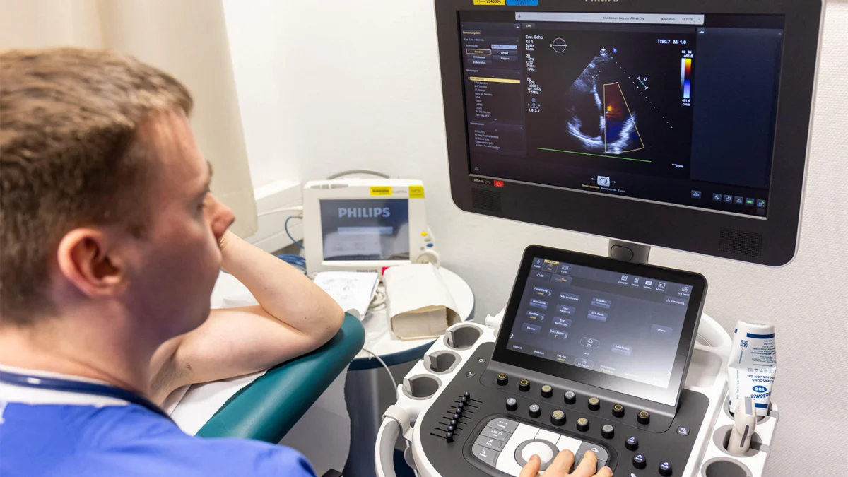
The AI-ECM is designed to operate within the existing workflow of clinical evaluations. Currently, patients suspected of having CA undergo echocardiography, followed by blood and urine tests. The AI-ECM aggregates this existing data, effectively acting as an intelligent assistant that flags patients who warrant more advanced, specialized testing, such as pyrophosphate imaging or biopsies.
Scalability and Global Health
Because the registry used for training the model was international and multiethnic, the AI-ECM offers a higher degree of generalizability than models trained on single-center, homogenous populations. This scalability suggests that the tool could be deployed in diverse healthcare settings, from high-resource academic medical centers to community hospitals, helping to standardize the quality of care for rare and underdiagnosed diseases.
Addressing the "Missed Diagnosis"
The primary implication of this research is the potential to reduce the time between symptom onset and diagnosis. Given that cardiac amyloidosis is increasingly treatable—especially when caught in the early stages—a shift from reactive to proactive diagnosis could translate into significant life-years gained for patients.
Future Directions
While the results are promising, Dr. Asch and his team acknowledge the need for prospective, external validation. Moving forward, the research community will likely focus on:
- Prospective Trials: Testing the AI-ECM in real-time clinical scenarios to observe how it impacts diagnostic turnaround times and patient outcomes.
- Surrogate Marker Development: Investigating whether the model can maintain its high sensitivity even when certain laboratory markers, like light-chain tests, are missing, thereby increasing its accessibility in resource-constrained environments.
- Integration into Clinical Decision Support Systems (CDSS): Collaborating with medical device manufacturers to embed the AI-ECM into diagnostic imaging hardware, making it a "plug-and-play" feature for clinicians worldwide.
In conclusion, the AI-ECM represents a sophisticated fusion of human clinical wisdom and machine learning capability. By transforming routine, often disconnected data points into a high-accuracy predictive model, this technology offers a robust solution to one of the most stubborn challenges in modern cardiology. As it moves toward prospective validation, the AI-ECM stands as a testament to the potential for AI to move beyond administrative tasks and into the core of clinical precision medicine.