


Featured Buzz – January 11, 2026
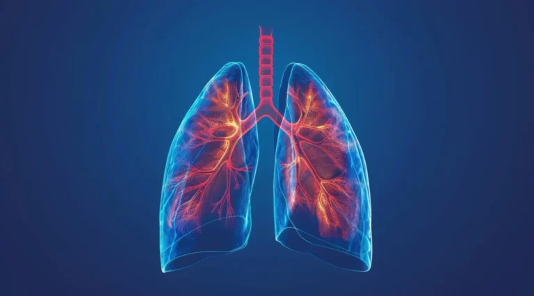
The landscape of pulmonary medicine is undergoing a profound transformation. As healthcare systems grapple with the dual challenges of diagnostic delays and the over-prescription of antibiotics, a new wave of research is leveraging artificial intelligence (AI) and point-of-care imaging to refine clinical precision. From the halls of Mount Sinai to the intensive care units of California, recent studies published in leading medical journals highlight a common theme: the integration of existing clinical data with machine learning is unlocking diagnostic potential that was previously hidden in plain sight.
1. ECGs as a Gateway to COPD Detection
The Diagnostic Dilemma
Chronic Obstructive Pulmonary Disease (COPD) remains one of the world’s most underdiagnosed conditions. Often characterized by persistent breathlessness and cough, it frequently evades detection until significant, irreversible lung damage has occurred. Currently, the gold standard for diagnosis is spirometry—a test that requires specialized equipment and patient effort, making it impractical for routine, asymptomatic screening.
Mount Sinai’s AI Breakthrough
Researchers at the Mount Sinai Health System have proposed a novel solution: repurposing the electrocardiogram (ECG). Because the heart and lungs share a tight physiological coupling, structural changes in the lungs caused by COPD often exert mechanical stress on the heart, leaving subtle electrical signatures on an ECG.
The research team undertook a massive data-mining effort, aggregating 208,231 ECGs from 18,225 patients with confirmed COPD, alongside a control group of 59,356 individuals matched for age, sex, and race. By feeding this vast dataset into a Convolutional Neural Network (CNN), the team trained the AI to recognize the specific patterns associated with COPD.
Implications for Early Intervention
The study, published in eBioMedicine, underscores that while an AI-enhanced ECG will not replace the definitive nature of spirometry, it serves as a powerful "early warning system." By identifying high-risk individuals during routine checkups, clinicians can initiate smoking cessation programs, prescribe targeted bronchodilators, and encourage pulmonary rehabilitation much earlier in the disease trajectory. This shift from reactive to proactive care could significantly reduce the long-term healthcare burden associated with advanced respiratory failure.
2. Precision in the NICU: Lung Ultrasound and Extubation
Predicting Success in Vulnerable Infants
For very low birth weight (VLBW) infants, the transition from mechanical ventilation to spontaneous breathing is a high-stakes clinical event. Failed extubation—when an infant requires reintubation within a week—is associated with higher mortality, longer hospital stays, and increased risk of neurodevelopmental impairment.
The University of Chicago Study
A study published in the Journal of Perinatology explored whether lung ultrasound (LUS) scores could act as a reliable predictive tool. The research followed 45 VLBW infants, tracking 53 extubation attempts. By performing an LUS three to six hours before the attempt, clinicians were able to assign a score reflecting the state of the neonatal lung.
The findings were definitive: the neonatal-adapted LUS score emerged as an exceptional predictor of extubation success. Furthermore, the study clarified the role of adjunctive therapies, noting that dexamethasone treatment did not lower LUS scores, providing clinicians with clearer guidelines on how to prepare their smallest patients for life without a ventilator.
Clinical Chronology and Methodology
- Patient Selection: 45 VLBW infants diagnosed with respiratory distress syndrome.
- Data Collection: Standardized LUS imaging conducted in the immediate pre-extubation window.
- Outcome Tracking: Reintubation rates monitored strictly within the seven-day post-extubation period.
- Validation: Correlation analysis confirmed that LUS scores accurately reflected the readiness of the infant’s lungs for independent ventilation.
3. Combating Antibiotic Overuse with AI-Driven Diagnostics
The Crisis of Inappropriate Antibiotic Use
In intensive care units, diagnosing lower respiratory tract infections (LRTIs) like pneumonia is notoriously difficult. Clinicians often face a "diagnostic gray zone" where they must choose between waiting for slow culture results or starting broad-spectrum antibiotics. The latter leads to the emergence of multi-drug resistant organisms.
The UCSF Diagnostic Model
Researchers at the University of California, San Francisco (UCSF), have developed an AI-integrated strategy that combines molecular biology with large language models (LLMs). The model utilizes a specific biomarker: FABP4, a gene found in lung fluid that modulates inflammation. When lung cells are healthy, FABP4 is highly expressed; when they are infected, expression drops.
By pairing this biological data with a generative AI analysis of the electronic medical record—specifically mining radiology reports and clinical notes—the researchers built a model that outperformed human ICU clinicians.
Official Responses and Performance Data
In an observational study, the AI model achieved a 96% diagnostic accuracy rate. When researchers compared the model’s performance against the actual clinical decisions made by ICU teams, they discovered that the AI’s adoption could have prevented over 80% of inappropriate antibiotic prescriptions.
"This study suggests that integrating a host biomarker with large language model analysis can improve LRTI diagnosis in critically ill adults," the authors stated in Nature Communications. The team is now pivoting toward validating this model for broader clinical use, with plans to expand the algorithm’s capabilities to identify sepsis—a move that could revolutionize how hospitals manage systemic infections.
4. Synthesis: The Future of Pulmonary Medicine
The common thread binding these three studies is the concept of "augmented clinical intelligence." Whether it is the Mount Sinai team using AI to "see" lung disease through heart-tracing equipment, or UCSF researchers using LLMs to synthesize complex clinical notes, the message is clear: technology is bridging the gap between raw data and actionable clinical wisdom.
Key Takeaways for Practitioners
- Early Screening: ECGs are no longer just for the heart; they are becoming viable tools for population-level COPD screening.
- Point-of-Care Reliability: Lung ultrasound is cementing its role as the superior bedside tool for managing neonatal respiratory transitions.
- Antibiotic Stewardship: AI models that synthesize biomarkers and unstructured clinical notes can provide the confidence needed to reduce unnecessary antibiotic use, thereby slowing the growth of antibiotic resistance.
Looking Ahead
The path forward for these technologies involves rigorous integration into the Electronic Health Record (EHR) ecosystem. The authors of the UCSF study have emphasized that their model is designed to be user-friendly, compatible with standard interfaces like GPT-4. As these tools move from the research paper to the patient bedside, they promise a future where diagnostic accuracy is higher, treatments are more targeted, and the overall quality of life for respiratory patients is significantly improved.
These advancements represent a pivotal moment in medicine. By embracing AI, the medical community is not replacing the clinician; rather, it is providing them with a more powerful lens through which to view patient health, ultimately turning data into life-saving action.
References and Further Reading:
- For details on the Mount Sinai COPD study, visit eBioMedicine.
- For the University of Chicago LUS findings, visit Journal of Perinatology.
- For the UCSF AI-LRTI diagnostic study, visit Nature Communications.