


In a groundbreaking development that promises to reshape how physicians monitor the long-term impact of psychological pressure, researchers at the Johns Hopkins University School of Medicine have successfully utilized deep learning artificial intelligence to identify the first biomarker of chronic stress visible on standard medical images.
The findings, set to be presented at the upcoming annual meeting of the Radiological Society of North America (RSNA), represent a paradigm shift in preventative medicine. By automating the measurement of adrenal gland volume through routine chest CT scans, the AI model provides a window into the physiological “wear and tear” caused by prolonged stress—a condition that has historically been notoriously difficult to quantify objectively.
The Hidden Burden: Chronic Stress as a Systemic Threat
Chronic stress is a ubiquitous modern ailment, yet it remains one of the most elusive health risks to track. According to the American Psychological Association, the impact of sustained stress extends far beyond emotional distress. It serves as a biological catalyst for a myriad of health issues, including hypertension, insomnia, muscle tension, and a suppressed immune system.
More critically, clinical literature has long linked the state of chronic stress to severe, life-altering conditions such as major depression, obesity, and cardiovascular disease. Until now, clinicians have relied on subjective tools—such as self-reported questionnaires—or cumbersome biological assessments, such as serial cortisol testing, which captures only a fleeting snapshot of the body’s hormonal state.
"Until now, we haven’t had a way to measure and quantify the cumulative effects of chronic stress, other than questionnaires or surrogate serum markers," explains Dr. Shadpour Demehri, professor of radiology at Johns Hopkins and senior author of the study. "For the first time, we can ‘see’ the long-term burden of stress inside the body using a scan that patients already receive every day in hospitals across the country."
Chronology of the Research
The journey to this discovery began with the recognition that tens of millions of chest CT scans are performed annually in the United States. Recognizing this vast, underutilized repository of health data, lead author Dr. Elena Ghotbi and her team set out to create a tool capable of mining this information for signs of systemic stress.
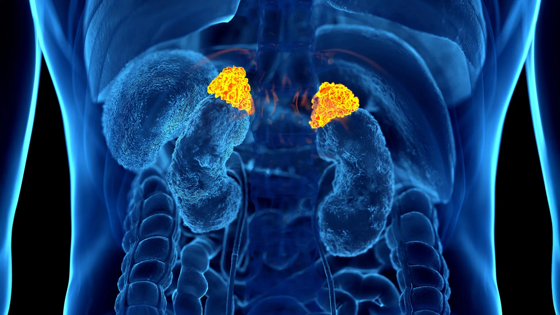
- Development of the Model: The research team developed a deep learning algorithm specifically trained to identify and segment the adrenal glands—the primary endocrine organs responsible for the body’s stress response—from existing CT datasets.
- Data Integration (2023–2024): The team analyzed data from 2,842 participants (average age 69.3 years) from the Multi-Ethnic Study of Atherosclerosis (MESA). This cohort was selected due to its comprehensive nature, which included not only imaging but also validated stress questionnaires, salivary cortisol measurements, and indicators of "allostatic load"—the total physiological burden accumulated over time.
- Refining the Metric: The researchers defined the "Adrenal Volume Index" (AVI), calculated as the adrenal volume in cubic centimeters divided by the height squared (cm³/m²).
- Validation: By comparing the AI-generated AVI against traditional markers (cortisol levels, blood pressure, glucose, and heart rate), the team confirmed that the adrenal size acts as a long-term "gauge" of chronic stress, effectively documenting the body’s physiological history in a way a single blood test cannot.
Supporting Data: Connecting Imaging to Clinical Outcomes
The study’s findings were statistically robust, linking higher AVI values directly to clinical indicators of poor health. The research demonstrated that individuals who reported higher levels of perceived stress consistently showed larger adrenal volumes.
Crucially, the study established a direct correlation between this imaging marker and cardiovascular health. High AVI values were linked to a higher left ventricular mass index—a structural change in the heart often associated with long-term cardiovascular strain. Perhaps most significantly, the team discovered that for every 1 cm³/m² increase in the Adrenal Volume Index, the risk of heart failure and mortality increased.
"With up to 10-year follow-up data on our participants, we were able to correlate AI-derived AVI with clinically meaningful and relevant outcomes," Dr. Ghotbi noted. "This is the very first imaging marker of chronic stress that has been validated and shown to have an independent impact on a cardiovascular outcome, namely, heart failure."
Official Responses and Expert Perspective
The medical community has greeted the findings with significant enthusiasm, noting that the "operationalization" of stress measurement could be a transformative tool in primary care.
Dr. Teresa E. Seeman, professor of epidemiology at UCLA and a pioneer in the study of stress and health, served as a co-author on the project. She emphasized the importance of the study’s practical application: "For over three decades, we’ve known that chronic stress can wear down the body across multiple systems. What makes this work so exciting is that it links a routinely obtained imaging feature with validated biological and psychological measures. It’s a true step forward in measuring the cumulative impact of stress on health."
Dr. Demehri added that the beauty of the system lies in its non-invasiveness. Because the adrenal glands are already captured in standard chest CT scans, this diagnostic insight requires no additional radiation, no extra appointments, and no extra costs to the patient. It essentially turns existing medical archives into a predictive health dashboard.
Implications for Future Clinical Practice
The implications of this technology are vast, particularly for middle-aged and older populations who frequently undergo routine imaging for unrelated screenings.
1. Risk Stratification
By integrating the AVI into standard radiology reports, doctors could gain a "stress score" for patients. This would allow for better cardiovascular risk stratification, helping cardiologists and primary care physicians identify high-risk individuals before they suffer a catastrophic event like a heart attack or stroke.
2. Preventive Intervention
If a patient’s CT scan reveals an elevated AVI, it could serve as a clinical "wake-up call." Physicians could use the data to initiate earlier interventions, such as lifestyle counseling, stress-management programs, or pharmaceutical management of blood pressure and inflammation, effectively closing the gap between psychological stress and physiological disease.
3. A New Standard in "Allostatic Load" Measurement
The ability to measure allostatic load—the wear and tear on the body—objectively is the "holy grail" of psychosomatic medicine. By using AI to distill complex hormonal, structural, and behavioral data into a single numerical index, the Johns Hopkins team has provided a language that both radiologists and primary care providers can use to communicate about the invisible toll of modern life.
4. Broadening the Scope
While the current study focused on cardiovascular outcomes, the researchers suggest that the potential applications are nearly limitless. As the model is refined, it could be used to study the correlation between adrenal volume and other stress-sensitive conditions, including cognitive decline, metabolic disorders, and immune dysfunction.
Conclusion
The development of the Adrenal Volume Index marks a historic moment in the intersection of artificial intelligence and preventive medicine. By translating the abstract concept of "chronic stress" into a tangible, measurable, and visual metric, researchers have bridged the divide between the mind and the body.
As the medical community prepares to digest these findings at the RSNA meeting, the focus will likely shift to how quickly this deep learning tool can be integrated into hospital software systems nationwide. In an era where the pace of life continues to accelerate, the ability to see the invisible consequences of that speed may be the most vital tool in the modern physician’s arsenal.
Research co-authors include Roham Hadidchi, Seyedhouman Seyedekrami, Quincy A. Hathaway, M.D., Ph.D., Michael Bancks, Nikhil Subhas, Matthew J. Budoff, M.D., David A. Bluemke, M.D., Ph.D., R. Graham Barr, and Joao A.C. Lima, M.D.