








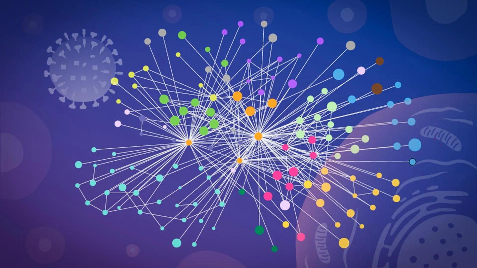
In a landmark study published in Nature Microbiology, an interdisciplinary team of researchers from EMBL Hamburg, the Leibniz Research Institute for Molecular Pharmacology (FMP), and the Charité – Universitätsmedizin Berlin has unveiled a high-resolution map of how the influenza A virus systematically commandeers human cells. By observing protein interactions in their native, intact cellular environments, the researchers have moved beyond traditional laboratory methods that rely on "breaking" cells, offering a revolutionary glimpse into the molecular mechanics of viral replication.
This breakthrough provides a vital roadmap for pharmaceutical developers, potentially paving the way for next-generation antiviral therapies and more effective seasonal vaccines. As influenza continues to claim between 3 and 5 million cases of severe illness annually—resulting in up to 650,000 deaths—the ability to visualize this "viral takeover" in real-time represents a significant leap forward in pandemic preparedness.
The Architecture of an Infection: How Viruses Hijack Host Machinery
To understand the scale of the researchers’ accomplishment, one must first appreciate the complexity of an influenza infection. Upon entering a human cell, the influenza A virus does not merely replicate; it initiates a total systemic rewrite of the host’s molecular operations. It releases its viral RNA, which carries the blueprints for producing viral proteins. These proteins then fan out across the cell, effectively hijacking the host’s organelles and metabolic pathways, transforming a healthy, functioning cell into a dedicated production line for new viral particles.
Historically, the difficulty in studying this process has been the "observation bias" inherent in traditional biochemistry. To measure protein-to-protein interactions, researchers have long relied on breaking cells apart. However, this destruction of the cellular environment often results in a "molecular collision" where proteins that never meet in a living cell are forced together in a test tube. Conversely, delicate, transient, or location-specific interactions—the very ones that drive the viral takeover—are often lost or distorted during the lysis process.
Chronology of a Breakthrough: From Concept to Computational Model
The success of this study was rooted in a collaborative effort that bridged the gap between advanced mass spectrometry and artificial intelligence.
Phase 1: Capturing the Moment (The XL-MS Innovation)
The research began when Jan Kosinski, Group Leader at EMBL Hamburg and the Centre for Structural Systems Biology (CSSB), sought a way to capture the "native context" of infection. The solution came through a partnership with Boris Bogdanow and Fan Liu at FMP Berlin. Together, they refined a specialized version of cross-linking mass spectrometry (XL-MS).
Unlike standard techniques, this tailored XL-MS approach allows scientists to "freeze" protein interactions while they are still occurring within an intact, living cell. By capturing these fleeting molecular handshakes, the team could identify exactly where and when the virus interacts with human host proteins during the replication cycle.
Phase 2: Integrating AlphaFold
Once the experimental data was captured, the team faced the daunting task of visualizing the structural geometry of these interactions. They turned to a modified version of AlphaFold, the Nobel Prize-winning protein structure prediction algorithm. By feeding their experimental cross-linking data directly into the model, the researchers provided the AI with physical constraints—data points confirming which parts of the viral and human proteins were in close proximity within the infected cell. This integration allowed the team to build highly accurate structural models of virus-host complexes that were previously thought to be too unstable or complex to predict.
Key Findings: The Two-Pronged Strategy of Influenza A
The study’s data revealed two primary strategies utilized by the influenza A virus to solidify its control over the host.
1. The Hemagglutinin Modification Pathway
The researchers focused on hemagglutinin, the viral surface protein responsible for initial cell attachment and entry. The team tracked this protein as it navigated the host’s internal transport and processing network. They discovered that the virus systematically recruits specific human proteins to assist in the proper folding and modification of hemagglutinin. Many of these human proteins were previously characterized by their "poorly understood" functions, suggesting that influenza has evolved to exploit cellular pathways that scientists have only just begun to map.
2. The Dissolution of Paraspeckles
Perhaps the most striking finding was the systematic destruction of "paraspeckles"—droplet-like compartments nestled within the cell nucleus. The team observed that the influenza virus consistently causes these structures to dissolve. This is no accident; as paraspeckles break apart, they release RNA-binding proteins that were previously sequestered. The virus appears to liberate these proteins to support its own replication. Furthermore, because paraspeckles are often involved in cellular stress responses and the regulation of antiviral genes, their destruction likely serves a dual purpose: fueling the virus while simultaneously crippling the cell’s primary defensive alarm system.
Official Perspectives: A New Standard for Virology
"Our work provides a new way to study flu-host interactions in their native context and with structural insight," said Jan Kosinski. He emphasized that the current findings represent a "snapshot" of the infection cycle, but more importantly, they provide a framework for future research. "It opens the door to studying flu-host interactions across the entire infection cycle, not just at one moment in time," he added.
Iuliia Kotova, the study’s first author, noted the profound nature of the paraspeckle discovery. "Watching these tiny organelles in the nucleus dissolve, consistently across every cell line and every flu strain we tested, told us this isn’t a side effect of infection—it might be a strategy," she explained.
From the analytical side, Boris Bogdanow, now at the Institute of Virology at Charité – Universitätsmedizin Berlin, highlighted the clinical potential: "XL-MS allows us to capture protein-protein interactions directly in infected intact cells, while also providing structural information… This gives us insight into the interface between the virus and the human cell and may, through structural modeling, help identify actionable targets for future pharmaceutical interventions."
Implications for Pandemic Preparedness
The collaborative nature of this study—spanning three major research hubs—has yielded a methodology that extends far beyond influenza A. By integrating cross-linking mass spectrometry, glycoproteomics, AlphaFold modeling, and high-end microscopy, the team has created a "modular" toolkit for virologists.
Broad Applicability
The researchers are optimistic that this "mapping in context" approach can be applied to a wide array of pathogens. While the specific host factors and viral proteins differ between diseases, the underlying strategy of combining experimental "in-cell" snapshots with AI-driven structural modeling is a universal template.
Future Research Directions
While this study utilized a laboratory-adapted strain of influenza, the team is already looking toward the future. The methodologies developed here are currently being evaluated for their ability to study viruses of greater pandemic concern, such as the highly pathogenic H5N1 avian influenza. By uncovering the specific interaction networks that allow these viruses to multiply, scientists hope to move from reactive vaccine development to proactive, target-specific drug design.
The ability to map these interactions at a structural level effectively turns the "black box" of viral infection into a transparent process. As the scientific community continues to refine these techniques, the era of "blind" drug discovery may soon be replaced by a new, precision-based era of antiviral medicine, where we do not just fight the virus—we dismantle its tools of sabotage before it even has the chance to start.