








Long before a human being draws their first breath, a silent, intricate construction project is underway. Within the protective confines of the womb, the human brain—the most complex structure in the known universe—is being meticulously assembled. At the helm of this biological marvel is a singular class of stem cells: radial glia.
For decades, these cells were viewed primarily as the scaffolding of the developing brain. Today, however, cutting-edge research from the University of California, Los Angeles (UCLA) is revealing that radial glia are far more than passive structural supports. They are dynamic "decision-makers," orchestrating the birth of the cerebral cortex—the seat of human thought, language, and memory—through complex metabolic processes and physical cellular "conversations."
Two landmark studies, published concurrently in the journals Cell and Science, have illuminated these hidden mechanisms, offering a new blueprint for understanding how the human brain achieves its unique size and complexity, and why, when these processes go awry, neurodevelopmental disorders emerge.
The Master Architects: Understanding Radial Glia
Radial glia are the biological engines of the developing cerebral cortex. These specialized stem cells possess a remarkable ability to divide and differentiate, spawning the vast array of neurons and support cells that define our cognitive capacity.
In humans, the cerebral cortex has undergone a massive expansion compared to other species, a phenomenon largely attributed to the prolonged and specialized activity of these radial glial cells. While they largely disappear by the time of birth, their biological legacy is profound. Furthermore, scientists have observed that similar cells occasionally "reawaken" in the context of brain cancers, suggesting that these ancient developmental programs hold keys to both the origins of human intelligence and the mechanisms of malignancy.
"Radial glia are the coolest cells that have ever existed," says Aparna Bhaduri, an assistant professor of biological chemistry at the David Geffen School of Medicine at UCLA. "They’re really key to making us human. But they’re also at the center of many neurodevelopmental and neuropsychiatric disorders, as well as cancer—so understanding how they make their decisions is one way to start understanding how those conditions arise."
Chronology of Discovery: A New Perspective on Development
For years, the scientific community operated under the assumption that brain development followed a rigid, pre-programmed genetic script. While genetics certainly provide the foundation, the UCLA research team has uncovered a far more nuanced reality. The findings suggest that development is a constant, real-time negotiation between the cell and its environment.
Phase 1: Mapping the Metabolic Engine
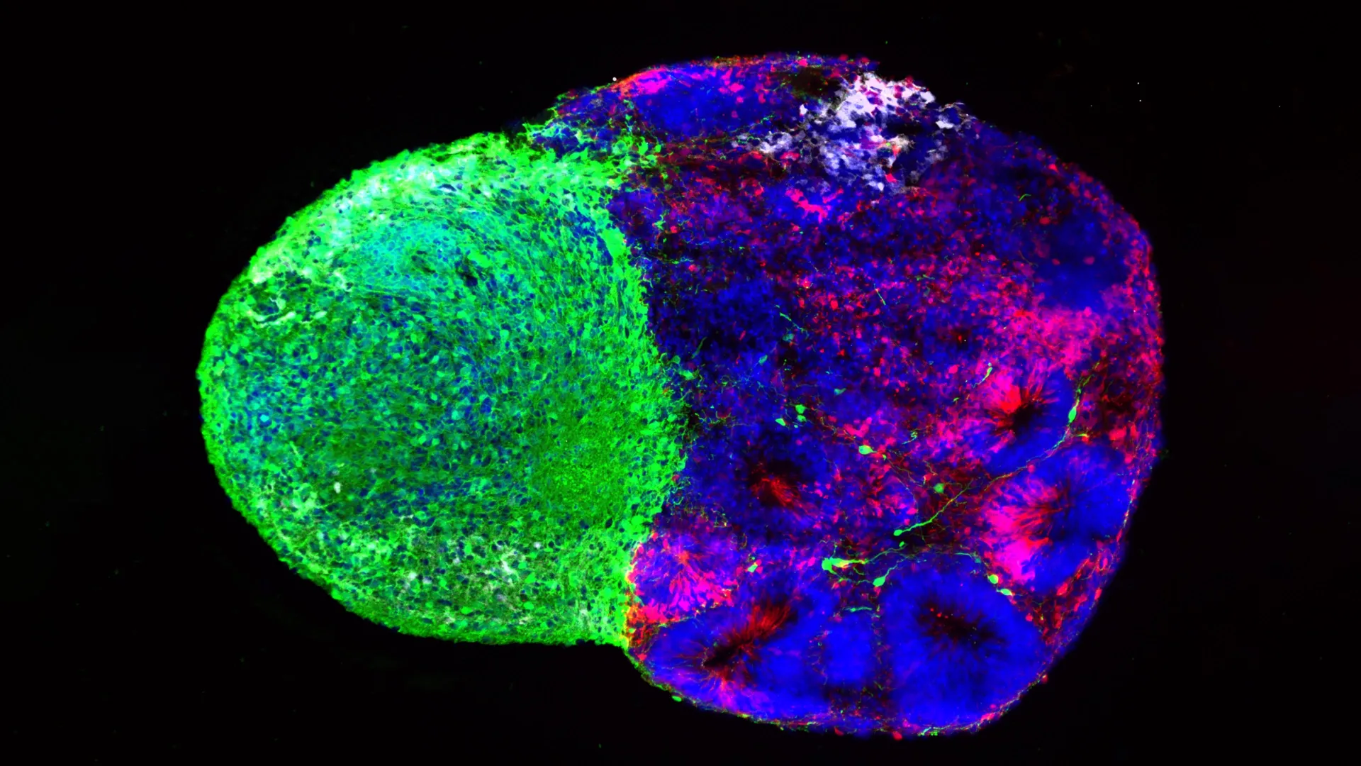
In the study published in Cell, the team, led by co-first authors Jessenya Mil and Jose Soto and in collaboration with the lab of Heather Christofk, sought to map the metabolic landscape of the developing human cortex. Using donated human tissue and sophisticated brain organoids—miniature, lab-grown versions of brain structures—they discovered that metabolism is not a passive bystander.
The researchers identified that radial glia rely heavily on the "pentose phosphate pathway." This specific metabolic route allows cells to efficiently convert glucose into the raw materials required for rapid cell division. When the researchers experimentally restricted glucose levels or inhibited this metabolic pathway, the radial glia underwent a dramatic shift in behavior. Instead of producing their standard output, they began generating inhibitory neurons—cells that typically appear much later in the developmental sequence.
This suggests that metabolic shifts are not merely consequences of development, but active regulators of it.
Phase 2: The Physical "Handshake" from the Thalamus
While the first study examined internal chemical processes, the second study, published in Science and led by Claudia Nguyen, investigated external physical inputs. The focus was on the thalamus, a deep-brain structure responsible for relaying sensory information.
It has long been known that the thalamus sends projections toward the cortex. What surprised researchers was the timing: these fibers arrive in the cortex far earlier than previously thought, long before their final synaptic connections are functional. The study revealed that these early-arriving fibers make direct physical contact with radial glia.
This contact acts as a "trigger," signaling the stem cells to increase the production of excitatory neurons—the primary information-carriers of the cortex. This interaction is particularly critical for the development of upper-layer neurons, the very cells that are most expanded in the human brain compared to rodents. This "physical handshake" appears to be a uniquely human developmental milestone.
Supporting Data and Technical Breakthroughs
The precision of these findings is largely due to the evolution of organoid technology. A decade ago, scientists were limited to animal models, which, while useful, fail to replicate the unique expansion and complexity of the human cortex.
The Role of Organoids and Assembloids
By utilizing "assembloids"—complex, multi-region organoids that mimic the interaction between different parts of the brain—the UCLA team could observe the direct physical interaction between thalamic projections and radial glia.
Key metrics from the research included:
- Metabolic Flux: Identifying that the pentose phosphate pathway is the primary metabolic driver in early radial glia.
- Cellular Output: Quantitative evidence showing a shift in neuron types (inhibitory vs. excitatory) based on metabolic and physical interventions.
- Genetic Association: Identifying the gene NRXN1 as a mediator of the thalamic-radial glial connection.
The researchers tested these interactions using patient-derived cells carrying a mutation in the NRXN1 gene, which is strongly associated with autism spectrum disorder. The altered thalamic signals in these models led to a disrupted balance between stem cell maintenance and neuronal production, providing a mechanistic link between early-stage cellular "conversations" and later neurodevelopmental outcomes.
Official Responses and Scientific Significance
Dr. Aparna Bhaduri, a member of the UCLA Broad Stem Cell Research Center and the UCLA Health Jonsson Comprehensive Cancer Center, emphasizes that these findings represent a fundamental shift in how we view the "developing" brain.
"What was surprising is that metabolism isn’t just a passive thing that happens in the background," says Dr. Bhaduri. "It can really control how stem cells make decisions."
The research community views these studies as a significant leap forward. By moving away from a purely genetic determinist model and toward one that integrates environmental, metabolic, and physical factors, scientists now have a more comprehensive toolkit to investigate:
- Maternal Nutrition: How metabolic shifts during pregnancy may influence fetal brain development.
- Disease Vulnerability: How mutations in genes like NRXN1 cause the "miscommunication" that leads to neuropsychiatric conditions.
- Cancer Research: Why dormant stem-cell programs (like those of radial glia) are reactivated in brain tumors.
Implications for the Future of Medicine
The implications of this research are broad and potentially transformative.
Understanding Neurodevelopmental Disorders
By pinpointing the exact moment when physical contact or metabolic signaling is interrupted, researchers may one day be able to identify biomarkers for autism, schizophrenia, and other neurodevelopmental disorders long before symptoms appear. Understanding the "decisions" these cells make is the first step toward intervening when those decisions go wrong.
The "Under the Hood" Approach
The shift in perspective—viewing metabolism and cellular contact as active drivers of development—is perhaps the most lasting contribution of these studies. It encourages future researchers to look past the DNA sequence and investigate the cellular environment. As organoid and assembloid technologies continue to improve, the ability to "peer under the hood" of human brain development will only sharpen.
A Path Toward Regenerative Medicine
Finally, while these studies focus on the developing brain, the insights into radial glia may hold secrets for regenerative medicine. If scientists can understand how these stem cells are instructed to produce specific types of neurons, it may eventually lead to therapies that "re-instruct" cells to repair damaged brain tissue in adults, potentially offering hope for conditions such as stroke, traumatic brain injury, or neurodegenerative disease.
As Dr. Bhaduri concludes, "Ultimately, these studies give us a glimpse under the hood of how these cells make decisions. Understanding those decisions is a first step toward understanding normal brain development, disease vulnerability and, potentially, how similar stem-cell programs operate in brain cancer."
This research was supported by the National Institutes of Health, the National Science Foundation, the Brain & Behavior Research Foundation, the Alfred P. Sloan Foundation, the Rose Hills Foundation, the Esther A. & Joseph Klingenstein Fund, the Simons Foundation, the Chan Zuckerberg Initiative, the NIH BRAIN Initiative Cell Atlas Network, the International Foundation for Ethical Research, the UCLA Broad Stem Cell Research Center’s Stem Cell Research Training Program, and the UCLA Health Jonsson Comprehensive Cancer Center and UCLA Broad Stem Cell Research Center Ablon Scholars Program.