









By Subramaniam C. Krishnan
September 28, 2026
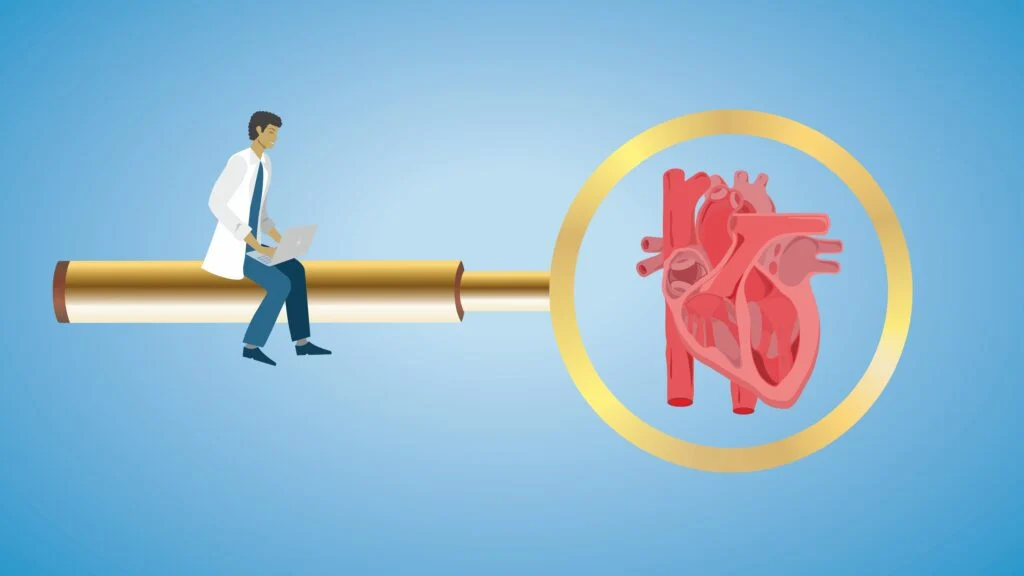
For more than a century, modern medicine has operated under a collective delusion: the belief that we have already mapped the final frontiers of human cardiac anatomy. As medical students, we are taught to memorize the heart’s chambers, valves, and vessels with the finality of a completed atlas. As surgeons, we navigate its architecture with the confidence of those walking through a well-lit room. As cardiologists, we interpret echocardiograms and MRIs so familiar that they have become nearly invisible to our trained eyes.
We treat anatomy as history—a chapter authored by the titans of the Renaissance, such as Andreas Vesalius, and solidified by Henry Gray’s legendary texts. We are told that the true frontiers of science lie in the abstract realms of genomics, artificial intelligence, and precision medicine. But what if we are wrong? What if the most significant medical secrets are not buried in our DNA, but hiding in plain sight within an organ we believed we had already mastered?
The Persistence of Observation: A Chronology of Discovery
My own journey toward this realization began in 2004, far from the gleaming high-tech labs of Silicon Valley. I found myself at the Jesse E. Edwards Registry of Cardiac Disease in St. Paul, Minnesota. Dr. Jesse E. Edwards, a luminary in cardiac pathology, possessed a rare foresight; he understood that today’s "normal" specimen could become tomorrow’s clinical enigma. He maintained a vast collection of preserved human hearts, treating careful observation as a legitimate—and vital—engine of discovery.
While studying these specimens on my own time, I encountered a peculiar configuration within the atrial septum that I had never encountered in any textbook. It was a pouch, opening into the left atrium, approximately 8 to 11 millimeters deep. It was not a hole, nor a valve, but a distinct, recessed structure.
My colleague and I launched a systematic investigation, scouring the registry’s extensive collection. In 2010, we published the first formal description of what we termed the "left atrial septal pouch" (LASP). The discovery was met with a mixture of professional intrigue and skepticism. How could a structure that exists in approximately one-third of the adult population remain formally unrecognized until the 21st century?
The answer is humbling: discovery is often limited not by a lack of advanced technology, but by our own narrow assumptions about where to stop looking. We had been looking at the heart, but we had stopped seeing it.
The LASP and the Clinical Implications of Anatomy
The left atrial septal pouch is more than a biological curiosity; it is a prime example of why anatomy remains a vital clinical science. In the biological hierarchy, development creates anatomy, which in turn determines local physiology. When that physiology is disrupted or challenged, it evolves into pathology.
The LASP is closely related to a patent foramen ovale (PFO)—a common "hole in the heart." However, the pouch functions differently. It resembles the left atrial appendage, which is well-known to cardiologists as a blind-ended cul-de-sac where blood can stagnate and eventually form clots. The danger of the LASP lies in its capacity for stasis. Unlike the left atrial appendage, where atrial fibrillation is often a prerequisite for clot formation, the LASP can facilitate stagnation and thrombus formation even in a patient with a perfectly normal heart rhythm.
Over the last 16 years, observational studies have consistently suggested that this anatomical variant may be a missing link in the mystery of "cryptogenic" strokes—strokes that occur without a clear, identifiable cause. For years, we could establish an association, but correlation is not causation. We needed to see if intervening in this structure could actually change patient outcomes.
Bridging the Gap: From Observation to Randomized Trials
The path from an anatomical curiosity to a clinical target is fraught with skepticism, a trajectory familiar to anyone who followed the history of PFO closure. For decades, the PFO was recognized by anatomists but dismissed as clinically irrelevant by many in the medical establishment. It took years of epidemiological data, refined imaging, and rigorous randomized trials to establish PFO closure as a gold-standard therapy for specific stroke populations.
The LASP appears to be following this exact path, albeit at a significantly accelerated pace. Recently, investigators at the Fuwai Hospital in Beijing broke new ground by reporting the first randomized trial evaluating the transcatheter elimination of the pouch. Using a septal occluder device originally designed for PFO closure, researchers demonstrated that mechanically excluding the pouch is not only feasible but is associated with a marked reduction in recurrent, MRI-detected ischemic brain lesions compared to medical therapy alone.
While no single trial should be viewed as the final word, this was a watershed moment: it was the first time we demonstrated that directly addressing this anatomical structure could fundamentally improve patient lives.
Scientific Evolution: Learning from Nature’s Design
The story of the LASP has also provided a roadmap for innovation. My subsequent work on an implant-free strategy to close PFOs grew directly out of my study of the pouch. We realized that a LASP can be understood as an incompletely fused PFO tunnel. This insight suggested that nature does not necessarily require complete adhesion across the entire overlap zone to achieve a seal.
If failed postnatal fusion creates a PFO, perhaps we could replicate the biological conditions that normally complete that fusion, inducing focal adhesions so the tunnel closes without the need for a permanent, foreign implant. This is the beauty of the scientific process: an anatomical observation leads to a physiological question, which in turn generates a therapeutic hypothesis. It is a slow, methodical engine of discovery that remains one of medicine’s most reliable tools.
The Future of Cardiac Medicine: A Balanced Approach
Modern medicine is currently obsessed with the "omics"—genomics, proteomics, and the burgeoning power of artificial intelligence. While these fields deserve the billions of dollars in investment they receive, they should not convince us that the map of the human body is complete.
As we pour resources into teaching computers to recognize patterns within the human body, we must be careful not to lose the innate, human ability to recognize them ourselves. The discovery of the left atrial septal pouch serves as a potent reminder that anatomy remains a living science. It teaches us that curiosity is not an outdated methodology, but a fundamental requirement for progress.
The next great medical breakthrough may not be hiding behind a genomic sequence or buried in a massive data set. It may be hiding in plain sight, tucked away in the architecture of an organ we thought we already knew. As we move forward into the next decade of cardiac research, we must continue to marry the technological prowess of the future with the foundational, observant rigor of the past.
When we look at the heart, we must look beyond the familiar silhouettes of the chambers and valves. We must look for the nuances, the pouches, and the hidden pathways that defy our current models. In the end, the most sophisticated machine we have is still the one beating within the chest of our patients—and it still has stories to tell if we are willing to listen.