










For decades, patients living with hypermobile Ehlers-Danlos syndrome (hEDS) have navigated a medical landscape characterized by skepticism. They report burning sensations, erratic heart rates, and a pervasive, whole-body sensitivity that defies the limitations of their joint-focused diagnosis. When standard neurological scans return "normal," patients are often left without answers, leading to a profound sense of medical gaslighting.
However, a landmark study recently published in Scientific Reports is fundamentally shifting the narrative. By peering beneath the skin of hEDS patients, researchers have uncovered evidence that the neurological symptoms associated with this condition are not only real but are distinct, measurable, and systemic.
Main Facts: The Intersection of Connective Tissue and Nerves
The study, led by a team of researchers in Italy, provides the most compelling evidence to date that Small Fiber Neuropathy (SFN) is a hallmark feature of hEDS. SFN is a condition involving damage to the small, unmyelinated nerve fibers that transmit pain and temperature sensations, while also regulating the autonomic nervous system—the "autopilot" of the body responsible for heart rate, blood pressure, and digestion.
While large nerve fibers govern movement and coordination, the small fibers are the body’s primary sensory and regulatory interface. In hEDS, these fibers appear to be under constant assault. The study compared 35 patients with hEDS-associated SFN against 38 patients with idiopathic SFN (iSFN)—a group with similar symptoms but no known underlying connective tissue disorder. The findings reveal that hEDS patients suffer from a unique, more aggressive neurological profile that distinguishes their condition from other forms of peripheral neuropathy.
A Chronology of Suffering: The Long Road to Diagnosis
One of the most startling revelations of the research is the stark disparity in symptom onset between the two groups.
- The Early Onset Gap: Patients with hEDS/SFN reported the first signs of nerve involvement at an average age of 19. In contrast, those with idiopathic SFN did not report symptoms until age 35.
- A Decade of Uncertainty: This 16-year discrepancy suggests that individuals with hEDS are living with unrecognized, untreated neurological damage for years, potentially decades, before receiving a formal diagnosis.
- The Burden of Duration: By the time these patients reach a clinic, the disease duration is significantly longer than in other neuropathy patients. This prolonged period of undiagnosed symptoms highlights a systemic failure in current diagnostic protocols, where connective tissue laxity is treated, but the secondary, often more debilitating, neurological damage is ignored.
Supporting Data: Mapping the Autonomic Disruption
The study employed a "comprehensive battery" of assessments, including sensory testing, autonomic function analysis, and, crucially, skin punch biopsies. The data collected provides a roadmap of the systemic chaos inherent in hEDS.
The Autonomic Crisis
The autonomic symptoms in the hEDS/SFN group were not just present; they were severe and pervasive. Patients reported high rates of:
- Gastrointestinal Distress: Chronic reflux, slow motility, and alternating bowel habits.
- Thermographic Dysfunction: Significant heat intolerance and dry skin, linked to impaired sweat gland innervation.
- The POTS Connection: Perhaps the most significant finding was the prevalence of Postural Orthostatic Tachycardia Syndrome (POTS). While 51.5% of the hEDS/SFN group met the criteria for POTS, the idiopathic group showed zero cases.
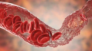
This finding challenges the long-held medical consensus that POTS in hEDS is solely due to "stretchy" blood vessels. Instead, the data points to a primary neurological failure: the nerve fibers themselves are failing to regulate the heart and blood pressure, suggesting that POTS in these patients is, at least in part, a neurogenic disorder.
Microscopic Evidence: The Skin Biopsy
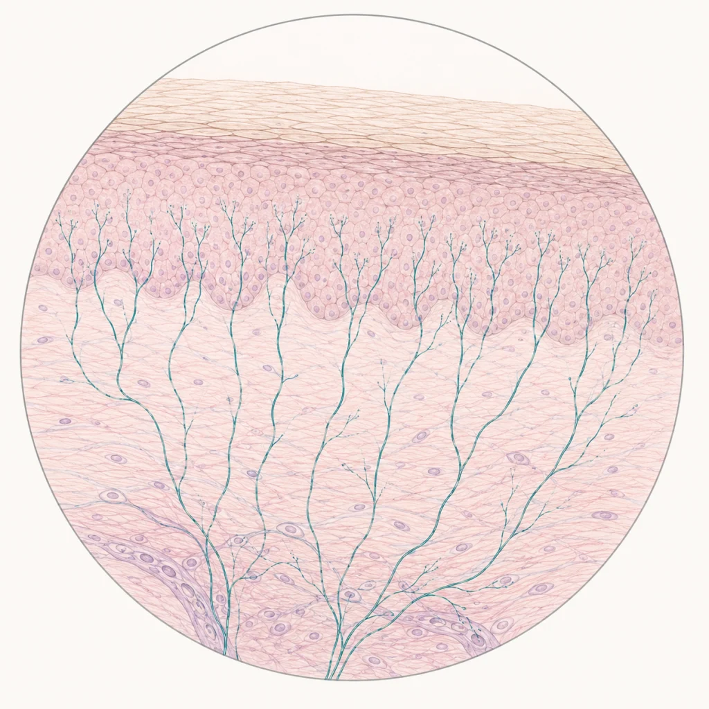
Skin biopsy is the gold standard for diagnosing SFN, involving the removal of a tiny sample of skin to count nerve endings. The biopsy results in this study were revealing:
- Greater Autonomic Loss: While sensory fiber density was reduced in both groups, the hEDS/SFN group exhibited a 2.5-fold higher loss of autonomic fibers.
- The Repair Paradox: Researchers identified a unique pattern of "alternating degeneration and regeneration." In hEDS patients, the nervous system appears to be caught in a cycle of constant damage and failed repair. This attempt at regeneration may actually contribute to the characteristic "burning" and "stinging" pain reported by patients, as misfiring, regrowing nerves are notoriously hypersensitive.
Official Perspectives and Clinical Implications
The implications for the medical community are profound. For years, clinicians have been trained to view hEDS as a joint-hypermobility disorder. This study argues that it must be reclassified—or at least approached—as a neuro-connective disorder.
"The pain and autonomic symptoms that so many people with hEDS experience are not only real, they are measurable," notes Dr. Jacqueline Moltzau Anderson, who highlights the necessity for a shift in standard care.
The Call for New Standards
The authors of the study strongly advocate for the integration of autonomic testing and skin biopsies into the standard diagnostic workup for hEDS. Currently, many patients are told their pain is "in their head" because standard MRIs and EMG (electromyography) tests—which only check large nerve fibers—come back normal. By incorporating specialized testing for small fibers, physicians can provide objective validation for a patient’s symptoms, preventing years of diagnostic delay.
Implications for Future Treatment
This research opens doors for more targeted therapeutic interventions. If the pain in hEDS is driven by a cycle of autonomic nerve fiber damage and attempted regeneration, the current "one-size-fits-all" approach to pain management—often relying on NSAIDs or physical therapy—is insufficient.
- Targeted Neurological Care: Patients with confirmed SFN could be candidates for therapies that specifically target nerve health, such as specialized supplements or medications that stabilize nerve membranes.
- Multidisciplinary Management: The data underscores that hEDS is a multi-system condition. Optimal care requires a team approach, integrating rheumatology (for joint laxity) with neurology (for nerve fiber health) and cardiology/autonomic specialists (for POTS and GI management).
- Validation as Medicine: For the patient community, the "biopsychosocial" model often used to dismiss chronic pain is dealt a blow. When a patient can point to a biopsy slide showing nerve fiber density, the conversation shifts from "is the pain real?" to "how can we treat the underlying nerve damage?"
Conclusion: A New Frontier in hEDS Care
The research published in Scientific Reports represents a paradigm shift. By quantifying the nerve damage associated with hEDS, the study provides a vital bridge between the subjective experience of chronic pain and the objective reality of biology.
For the millions of people living with hEDS, this study is a long-overdue validation. It confirms that the burning, the dizziness, the gut issues, and the fatigue are not just "symptoms of hypermobility"—they are the manifestations of a complex, damaged nervous system. Moving forward, the medical community must adopt these findings to move beyond simply managing joint dislocations and toward a more comprehensive, compassionate, and science-based approach to the neurological health of hEDS patients.
The goal now is clear: to ensure that the "normal" results on a standard neurological exam are no longer a barrier to care, but a prompt to look deeper—right down to the nerve fibers beneath the skin.