


For decades, the study of human vascular health has been constrained by a fundamental disconnect between reality and the laboratory. Human blood vessels are dynamic, organic structures characterized by intricate branching, unpredictable curvatures, and varying diameters. Yet, for years, the standard approach in biomedical engineering was to model these vessels as simple, straight, uniform tubes. While these simplified designs provided a baseline for understanding basic fluid dynamics, they failed to capture the chaotic, high-stakes environment where life-threatening vascular diseases—such as aneurysms and stenoses—actually manifest.
Researchers at the Texas A&M University Department of Biomedical Engineering are now bridging this gap. By developing a highly customizable, "organ-on-a-chip" vessel system, the team is enabling scientists to simulate the complex, non-linear architecture of the human circulatory system. This breakthrough offers a powerful new platform for disease modeling, personalized medicine, and non-animal pharmaceutical testing.
The Problem with Simplistic Models
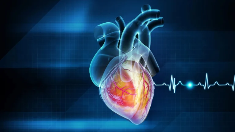
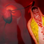
The human vascular system is far from a collection of smooth pipes. It is a labyrinth of physiological transitions. When a vessel branches, widens into an aneurysm, or narrows due to stenosis, the physical forces acting upon the vessel walls—specifically, shear stress—change dramatically. These mechanical forces are not merely background noise; they are active signals that govern cellular health, inflammation, and the progression of disease.
"There are branched vessels, or aneurysms that have sudden expansion, and then stenosis that restricts the vessel," explains Jennifer Lee, a master’s student in biomedical engineering who spearheaded the design of the new vessel-chip. "All these different types of vessels cause the blood flow pattern to be significantly changed, and the inside of the blood vessel is affected by the level of shear stress caused by these flow patterns. That’s what we wanted to model."
By ignoring these architectural nuances, previous models often missed the very conditions that lead to cardiovascular complications, rendering many drug-testing results less predictive of human outcomes.
Chronology of an Innovation: From Undergraduate Curiosity to Published Science
The journey toward this advanced vessel-chip began within the Bioinspired Translational Microsystems Laboratory, led by Dr. Abhishek Jain, an associate professor and the Barbara and Ralph Cox ’53 faculty fellow in biomedical engineering.
The lab’s trajectory toward this breakthrough can be traced back several years. Initial efforts were centered on proving the feasibility of organ-on-a-chip technology, with former graduate student Dr. Tanmay Mathur successfully developing a foundational straight vessel-chip design. This early work proved that microfluidic devices could effectively replicate the environment of a blood vessel. However, both Dr. Jain and his team recognized that a straight tube was only the beginning.
The Path to Discovery
Jennifer Lee entered the lab as an undergraduate honors student seeking a departure from traditional coursework. With little prior exposure to the complexities of organ-on-a-chip technology, she underwent a rapid immersion in the field. Her aptitude for the work was immediate, and she soon transitioned into the Master of Science fast-track program at Texas A&M, allowing her to commit to the long-term, high-risk research required to push the project forward.
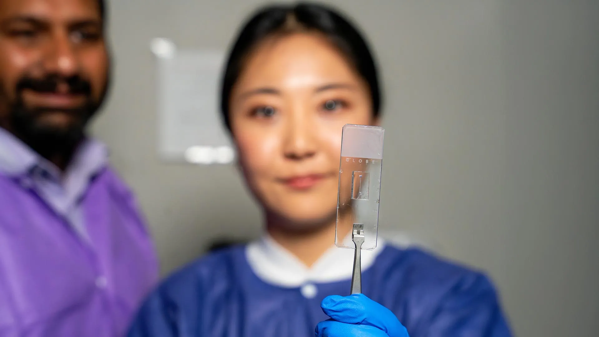
Her dedication culminated in a sophisticated design capable of replicating the wide range of physiological shapes seen in human anatomy. This achievement was recently validated by the scientific community: Lee’s research was published in the prestigious journal Lab on a Chip, and her work will be featured on the cover of the journal’s May 2025 issue.
Supporting Data and Technical Significance
The vessel-chip is a microfluidic device that operates at the micron scale, allowing for the precise control of fluid flow and cellular placement. Unlike traditional cell cultures in Petri dishes, which are static, these chips are "living" environments.
The Power of "Living" Vessels
The true innovation of the Texas A&M model lies in its ability to support actual biological tissue. "We can now start learning about vascular disease in ways we’ve never been able to before," says Dr. Jain. "Not only can you make these structures complex, you can put actual cellular and tissue material inside them and make them living. These are the sites where vascular diseases tend to develop, so understanding them is critical."
Currently, the model utilizes endothelial cells—the delicate, single-layer lining that coats the interior of every blood vessel. These cells are highly sensitive to blood flow and are the primary mediators of vascular health. By placing these cells into curved or branched channels, the researchers can observe how they respond to varying levels of shear stress in real-time, providing a high-fidelity window into how disease begins at the cellular level.
Official Responses and Strategic Vision
The development of this technology is not just an academic achievement; it represents a strategic shift in how the medical community approaches drug development and regulatory approval. By providing a human-centric, non-animal testing platform, these chips align with a global push to reduce reliance on animal models, which are often costly, ethically complex, and biologically distinct from humans.
Expanding the "Fourth Dimension"
Dr. Jain describes the current stage of the research as only the beginning. The lab is already conceptualizing the next iteration of the technology. "We are progressing and creating what we call the fourth dimensionality of organs-on-a-chip," Jain explains. "Where we not only focus on the cells and the flow, but this interaction of cells and flow in more complex architectural states, which is a new direction in the field."
Future iterations of the chip will likely incorporate multiple cell types, such as smooth muscle cells and immune cells, to recreate the full, multi-layered thickness of a blood vessel wall. This will allow researchers to observe how systemic inflammatory responses—a hallmark of many vascular diseases—interact with mechanical stress to cause blockages or ruptures.
Broader Implications for Medicine
The implications of this technology are far-reaching:
- Personalized Medicine: Because the chips can be tailored to individual patients, doctors could theoretically use a patient’s own cells to create a "digital twin" of their vascular network. This would allow for the pre-testing of drugs to determine efficacy and safety before a single pill is ingested.
- Pharmaceutical Efficiency: The current model offers a more robust platform for pharmaceutical companies to screen drug candidates. By identifying failures earlier in the process, companies can save millions in development costs and accelerate the delivery of life-saving treatments to the market.
- Disease Modeling: Researchers can now replicate rare or complex vascular conditions in the lab, providing insights that were previously obscured by the limitations of traditional 2D culture models.
Mentorship and Professional Development
Beyond the technical findings, the project serves as a case study for the value of university research programs that emphasize high-impact, high-risk projects for students. Dr. Jain credits Lee’s success to a combination of technical rigor and personal resilience.
"Jennifer demonstrated perseverance, curiosity, and creativity," Jain notes. "Our fast-track program enables students like Jennifer to take on high-impact research and not just do a science project, but take it all the way to its outcome and get it published."
For Lee, the experience has been about more than just data collection. The collaborative nature of the Bioinspired Translational Microsystems Laboratory forced her to develop soft skills—teamwork, project management, and effective communication—that are essential for any career in the biomedical sciences. "It’s such a good environment to interact with not only peers but also graduate students and postdoctoral researchers," Lee reflects. "You’re able to learn teamwork and communication, work ethic, and just trying different things out."
Funding and Future Outlook
The scale and ambition of this research have attracted significant attention and financial support from a wide array of high-profile entities. The project has been bolstered by funding from the U.S. Army Medical Research Program, NASA, the Biomedical Advanced Research and Development Authority (BARDA), the National Institutes of Health (NIH), the U.S. Food and Drug Administration (FDA), the National Science Foundation (NSF), and the Texas A&M University Office of Innovation Translational Investment Funds.
As the team prepares for the May 2025 publication, the focus remains on scaling the complexity of the vessel-chip and ensuring its transition from a laboratory tool to a clinical asset. By proving that the human vascular system can be accurately represented in a micro-scale environment, the Texas A&M team is not only changing the shape of the vessels they study—they are changing the shape of medical research itself.