










For decades, the standard approach to studying human vascular biology in the laboratory has relied on a fundamental, yet inherently flawed, simplification: the straight, uniform tube. While these models have provided foundational insights into cardiovascular mechanics, they ignore the chaotic, beautiful, and often dangerous reality of the human circulatory system. Our blood vessels do not exist as static pipes; they branch, bifurcate, widen into aneurysms, and narrow into stenoses. These geometric complexities are not merely structural—they are the primary theaters where vascular diseases are born.
Now, a team of researchers at the Texas A&M University Department of Biomedical Engineering is dismantling this paradigm. By pioneering a customizable "vessel-chip" system, the Bioinspired Translational Microsystems Laboratory is moving beyond the constraints of traditional models, offering a high-fidelity window into how complex blood flow patterns dictate the progression of life-threatening diseases.
The Limitations of Conventional Models
The human vascular system is a masterwork of engineering, designed to transport life-sustaining oxygen and nutrients through thousands of miles of vessels. However, this system is prone to failure, particularly at junctions and irregular segments where fluid dynamics become turbulent. In traditional laboratory settings, researchers have largely utilized straight microchannels to simulate these pathways.
"While useful, those simplified designs failed to reflect the conditions where many vascular diseases actually develop," notes the research team at Texas A&M. When blood flow encounters a curve, a branch, or a constriction, it experiences changes in "shear stress"—the frictional force exerted by blood flow against the vessel walls. These forces are critical biological signals that determine whether endothelial cells—the gatekeepers lining our blood vessels—remain healthy or transition into a diseased state.
By failing to replicate these complex shapes, scientists have historically missed the nuances of how shear stress contributes to atherosclerosis, aneurysms, and other vascular pathologies.
Chronology of Innovation: From Straight Lines to Complex Architectures
The development of the new vessel-chip system is the culmination of years of iterative research within the Bioinspired Translational Microsystems Laboratory, led by Dr. Abhishek Jain, an associate professor and the Barbara and Ralph Cox ’53 Faculty Fellow in Biomedical Engineering.
The Foundation
The journey began with the work of Dr. Tanmay Mathur, a former graduate student under Dr. Jain. Several years ago, Mathur successfully developed a straight vessel-chip design. While seemingly simple, this served as the essential proof-of-concept, establishing that human cells could be cultured within microfluidic devices to simulate the basic physiological environment of a blood vessel.
The Leap in Complexity
The baton was eventually passed to Jennifer Lee, a master’s student who joined the lab as an undergraduate honors student. Recognizing that the "straight-tube" model was insufficient for capturing the mechanical triggers of disease, Lee set out to engineer a chip capable of reproducing the diverse, irregular geometries of the human body.
Her work, which focuses on replicating branched vessels, sudden expansions (aneurysms), and constrictions (stenosis), represents a significant leap forward. Published in the journal Lab on a Chip—and slated for the cover of the May 2025 issue—Lee’s research successfully demonstrates that we can now replicate these high-risk areas in a controlled, scalable environment.
The Mechanics of the Vessel-Chip
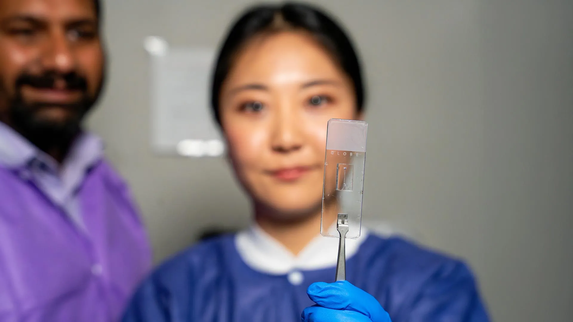
At its core, the vessel-chip is a marvel of microfluidics. These devices, often no larger than a thumb drive, utilize precise channels to replicate the scale of human microvasculature. By lining these chips with human endothelial cells, researchers create "living" vessels.
"There are branched vessels, or aneurysms that have sudden expansion, and then stenosis that restricts the vessel," explains Lee. "All these different types of vessels cause the blood flow pattern to be significantly changed, and the inside of the blood vessel is affected by the level of shear stress caused by these flow patterns. That’s what we wanted to model."
By controlling the geometry of the chip, the team can manipulate the flow dynamics to mimic the exact conditions found in a patient suffering from a specific vascular condition. This provides a platform for testing pharmaceutical interventions that is far more accurate than traditional animal models, which often fail to translate to human biology due to fundamental differences in cardiovascular architecture.
Official Responses and Strategic Implications
The implications of this technology extend far beyond the laboratory bench. Dr. Jain emphasizes that the ability to incorporate actual patient-derived cellular and tissue material makes these chips a game-changer for personalized medicine.
"We can now start learning about vascular disease in ways we’ve never been able to before," Dr. Jain says. "Not only can you make these structures complex, you can put actual cellular and tissue material inside them and make them living. These are the sites where vascular diseases tend to develop, so understanding them is critical."
The research has attracted significant interest from federal and national agencies, highlighting the strategic importance of this work. The project has received robust support from the U.S. Army Medical Research Program, NASA, the Biomedical Advanced Research and Development Authority (BARDA), the National Institutes of Health (NIH), the U.S. Food and Drug Administration (FDA), the National Science Foundation (NSF), and the Texas A&M University Office of Innovation Translational Investment Funds.
These partnerships reflect a shared understanding that moving away from animal testing toward high-fidelity, human-based "organ-on-a-chip" models is the future of drug discovery and safety testing.
Bridging the Gap: Education and Professional Development
The success of the vessel-chip project is as much a testament to Texas A&M’s pedagogical approach as it is to the engineering itself. Jennifer Lee’s progression from an undergraduate novice to a published researcher in the Lab on a Chip journal highlights the efficacy of the university’s fast-track master’s program.
Dr. Jain speaks highly of the culture fostered in his lab: "Jennifer demonstrated perseverance, curiosity, and creativity and started taking up research projects very quickly. Our fast-track program enables students like Jennifer to take on high-impact, high-risk research and not just do a science project, but take it all the way to its outcome and get it published."
For Lee, the experience was transformative, providing skills that transcend the technical requirements of biomedical engineering. "It’s such a good environment to interact with not only peers but also graduate students and postdoctoral researchers," Lee notes. "You’re able to learn teamwork and communication, work ethic, and just trying different things out. I think it’s such a valuable experience that students have available."
The Future: The "Fourth Dimension" of Micro-Physiology
Despite the success of the current model, the research team is not resting on its laurels. The current iteration focuses primarily on endothelial cells, but the team is already looking toward the next phase of development.
"We are progressing and creating what we call the fourth dimensionality of organs-on-a-chip," says Dr. Jain. "Where we not only focus on the cells and the flow, but this interaction of cells and flow in more complex architectural states, which is a new direction in the field."
Future iterations of the vessel-chip will likely incorporate multiple cell types—such as smooth muscle cells and immune cells—to create a more comprehensive representation of the vascular wall. By introducing these additional layers, researchers hope to observe how different tissues interact with one another and with the mechanical forces of blood flow in real-time.
Conclusion
The work emanating from Texas A&M’s Bioinspired Translational Microsystems Laboratory serves as a reminder that progress in medicine is often preceded by a shift in how we perceive the body itself. By moving beyond the "straight tube" assumption, Dr. Jain, Jennifer Lee, and their colleagues have opened the door to a deeper understanding of cardiovascular health.
As the field of organs-on-a-chip continues to mature, these complex, living architectures promise to revolutionize how we diagnose, treat, and ultimately prevent the vascular diseases that remain a leading cause of morbidity worldwide. With the backing of major federal agencies and a commitment to nurturing the next generation of scientific talent, the team at Texas A&M is positioned at the forefront of a new era in biomedical engineering—one where the complexity of the human body is finally met with the complexity of the tools used to study it.