











Featured Buzz – January 11, 2026
The landscape of pulmonary medicine is undergoing a profound transformation. As clinical demand rises for more efficient diagnostic tools, researchers are increasingly turning to the synergy between artificial intelligence (AI) and non-invasive imaging to bridge the gap between routine screening and definitive diagnosis. This week, three landmark studies—published in eBioMedicine, the Journal of Perinatology, and Nature Communications—illustrate how digital health and refined ultrasound techniques are enhancing the precision of care for conditions ranging from Chronic Obstructive Pulmonary Disease (COPD) to life-threatening lower respiratory tract infections (LRTIs) in the ICU.
1. AI-Powered ECGs: A New Frontier for COPD Screening
The Diagnostic Challenge
Chronic Obstructive Pulmonary Disease (COPD) remains one of the world’s leading causes of morbidity and mortality. Often underdiagnosed until the disease reaches an advanced stage, COPD requires timely intervention to mitigate irreversible lung damage. Currently, the gold standard for diagnosis is spirometry—a test that, while effective, is not always performed during routine clinical encounters, particularly in primary care settings where patients may present for unrelated cardiac issues.
The Mount Sinai Breakthrough
Researchers from the Mount Sinai Health System have introduced a novel methodology: leveraging the ubiquitous electrocardiogram (ECG) as a gateway to COPD detection. ECGs are standard practice for assessing cardiac health, making them a high-utility, low-cost screening tool if they can be repurposed to identify pulmonary pathology.
The research team curated a massive dataset consisting of 208,231 ECGs from 18,225 COPD patients and over 550,000 ECGs from age- and sex-matched controls. By training a Convolutional Neural Network (CNN) on this vast repository, the team enabled the model to recognize subtle electrical patterns in the heart that correlate with lung hyperinflation and the mechanical strain characteristic of COPD.
Implications for Clinical Practice
The study, published in eBioMedicine, suggests that while AI-interpreted ECGs will not supersede spirometry, they serve as a powerful "early warning system." By integrating this AI layer into existing clinical workflows, physicians could flag at-risk patients during routine check-ups. "Earlier recognition may facilitate timely smoking cessation, targeted therapies, and pulmonary rehabilitation," the authors noted, emphasizing that the reduction in the healthcare burden could be substantial if the disease is caught in its nascent stages.
2. Precision Pulmonology: Ultrasound Predicts Extubation Success in Infants
The Vulnerability of VLBW Infants
For very low birth weight (VLBW) infants, the transition from mechanical ventilation to independent breathing is a high-stakes clinical event. Failed extubation—the inability of an infant to maintain stable breathing after the removal of a ventilator tube—is associated with prolonged hospital stays, increased risk of infection, and long-term neurological complications.
Chronology of the University of Chicago Study
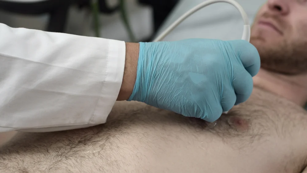
Researchers at the University of Chicago sought to refine the decision-making process for extubation by utilizing lung ultrasound (LUS) scores. The study focused on 45 VLBW infants suffering from respiratory distress syndrome.
- Baseline Assessment: 53 extubation attempts were monitored across the cohort.
- Real-time Monitoring: LUS examinations were conducted three to six hours prior to each planned extubation.
- Outcome Definition: A "failure" was strictly defined as any requirement for reintubation within seven days.
Supporting Data and Findings
The data indicated that the neonatal-adapted LUS score serves as a highly reliable predictive metric. Infants with higher ultrasound scores—reflecting improved lung aeration and reduced fluid accumulation—demonstrated significantly higher success rates in remaining off the ventilator. Furthermore, the study addressed a common clinical debate regarding medication, finding that dexamethasone treatment did not skew the diagnostic utility of the LUS score, reinforcing its reliability across diverse treatment protocols.
3. Combating Antibiotic Overuse with AI and Biomarkers
The Crisis of Inappropriate Antibiotic Use
In intensive care units (ICUs), the over-prescription of antibiotics is a global health crisis, fueling the rise of multidrug-resistant pathogens. Differentiating between a bacterial infection and non-infectious inflammation in critically ill patients with lower respiratory tract infections (LRTIs) is notoriously difficult.
A Multimodal Diagnostic Strategy
A research team from the University of California, San Francisco (UCSF), has developed a sophisticated diagnostic model that marries biological data with artificial intelligence. The approach relies on two pillars:
- Host Biomarkers: The model focuses on the gene FABP4, which is expressed in lung fluid. Researchers found that FABP4 levels are significantly suppressed in the presence of an actual bacterial infection compared to sterile inflammation.
- Generative AI Analysis: By feeding this biomarker data into a GPT-4 interface alongside clinical notes and radiology reports (chest X-rays), the model synthesizes a holistic view of the patient’s condition.
Official Results and Future Direction
In an observational study of critically ill adults, the AI-biomarker model achieved a diagnostic accuracy of 96%, significantly outperforming the intuition of seasoned ICU clinicians. The potential for impact is staggering: the study authors estimate that widespread adoption of this tool could reduce inappropriate antibiotic use by more than 80%.
"This study suggests that integrating a host biomarker with large language model analysis can improve LRTI diagnosis in critically ill adults," the researchers stated. The team is now pivoting toward validation for broader clinical deployment and exploring the potential of this diagnostic engine for the early identification of sepsis.
Synthesis: The Future of Digital Pulmonary Medicine
The confluence of these three studies highlights a broader trend: the digitization of diagnostics. Whether it is the repurposing of ECGs, the advancement of bedside ultrasound, or the use of large language models to interpret complex biological data, the message is clear—technology is making the invisible visible.
Implications for the Healthcare System
The implications of these advancements are manifold:
- Efficiency: By utilizing tools already present in the clinical environment (ECGs and standard medical records), healthcare systems can improve diagnostic speed without the need for expensive, specialized infrastructure.
- Accuracy: Reducing the reliance on subjective clinical judgment in favor of data-driven insights minimizes the risk of human error in high-stress environments like the ICU.
- Economic Impact: Earlier diagnosis of COPD and reduced reliance on inappropriate antibiotics not only improve patient outcomes but also lower the long-term financial burden on hospitals by reducing ICU days and readmission rates.
The Path Forward
As these models move from observational studies to clinical trials and eventual real-world application, the focus must remain on integration and safety. The UCSF team’s reliance on HIPAA-compliant interfaces for their GPT-4 model underscores the necessity of data privacy and ethical implementation in the AI era.
Ultimately, these innovations represent a paradigm shift. We are moving away from reactive medicine—where we treat symptoms only after they become severe—toward a proactive, data-informed model of care. As researchers continue to refine these algorithms, the standard of care for pulmonary health is poised to become safer, faster, and significantly more precise.
References:
- eBioMedicine (2025). AI-enhanced ECG interpretation for COPD screening.
- Journal of Perinatology (2025). Lung ultrasound as a predictor for extubation in VLBW infants.
- Nature Communications (2025). Multimodal AI diagnostics for LRTIs in critical care.