











Introduction: The Hidden War Within Our Cells
Every year, the seasonal influenza virus triggers between 3 and 5 million cases of severe illness globally, claiming up to 650,000 lives in the process. Beyond its seasonal burden, influenza A remains a perennial candidate for global pandemics, a legacy underscored by the devastating 1918 Spanish Flu. Despite its ubiquity, the exact molecular mechanisms by which this virus subverts human cellular machinery have long remained partially obscured.
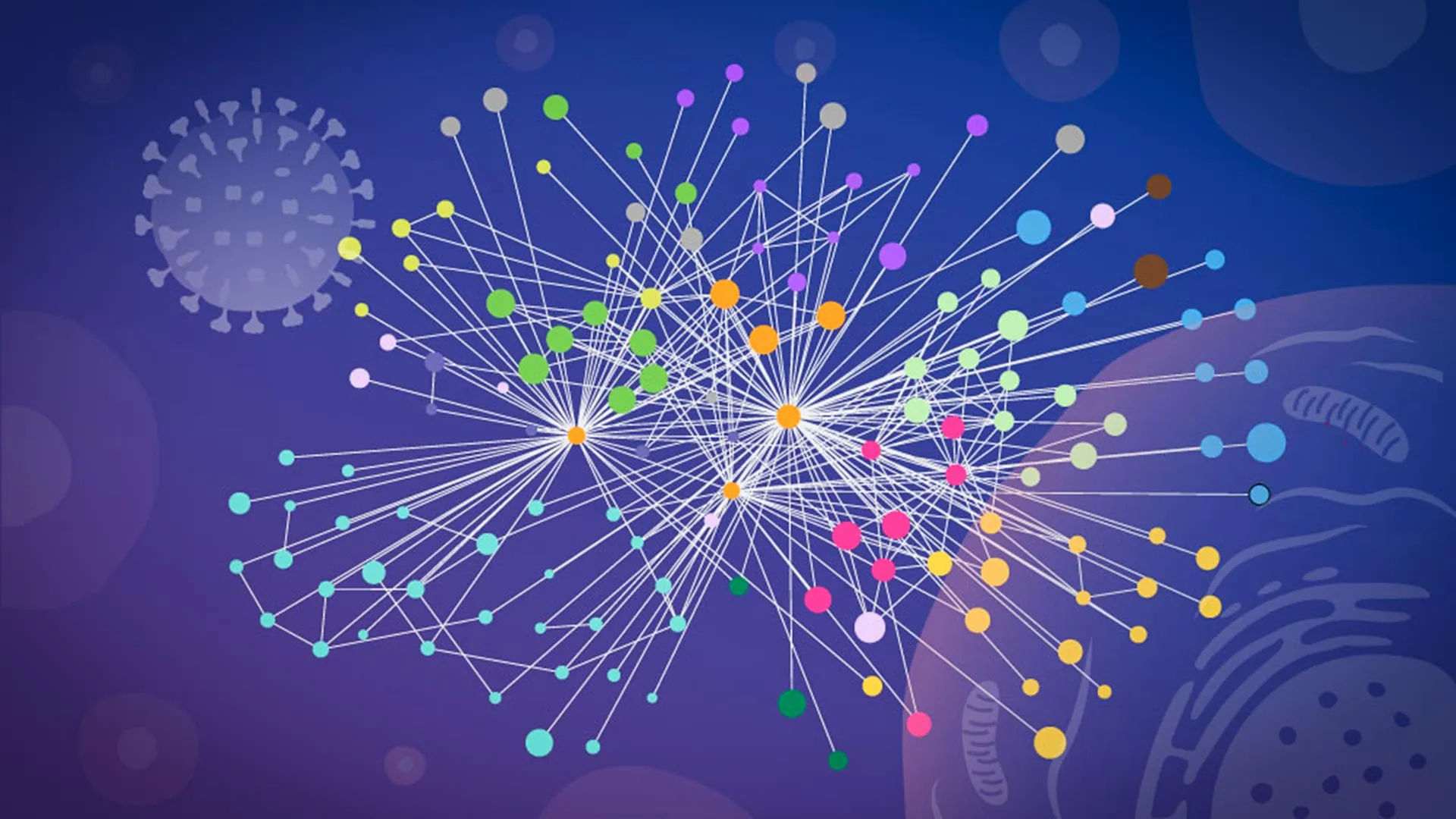
A landmark study published in Nature Microbiology has finally shed light on this biological insurgency. Researchers from EMBL Hamburg, in collaboration with the Leibniz Research Institute for Molecular Pharmacology (FMP) and Charité – Universitätsmedizin Berlin, have produced an unprecedented, high-resolution map of how influenza A reshapes infected human cells. By observing protein interactions directly within the native, intact environment of a living cell, the team has bypassed the limitations of traditional biochemical methods, revealing not just what the virus does, but how it orchestrates its takeover.
The Challenge of Observation: Breaking the "Broken-Cell" Barrier
For decades, the study of protein-protein interactions (PPIs) has relied on a trade-off. To understand how viral and host proteins communicate, scientists typically "lysed"—or broke open—infected cells to isolate proteins for analysis. However, this destruction of cellular architecture creates a significant experimental bias.
When a cell is ruptured, the internal environment is fundamentally altered. Proteins that were separated by membranes or located in different organelles suddenly collide in a soup of cellular debris, leading to "false-positive" interactions. Conversely, transient, weak, or highly localized interactions—often the most critical for viral replication—are frequently lost during the extraction process.
"Tracking protein-protein interactions during an active infection is extremely difficult," explains Jan Kosinski, Group Leader at EMBL Hamburg and the Centre for Structural Systems Biology (CSSB). "Many earlier studies depended on biochemical techniques that required scientists to break cells open. That process can distort what was happening inside the living cell."
To overcome this, the research team turned to a specialized version of cross-linking mass spectrometry (XL-MS). This technique involves using chemical "fixers" to lock proteins into their interacting positions before the cell is broken apart. Developed by collaborators Boris Bogdanow and Fan Liu at FMP Berlin, this tailored workflow allowed the researchers to capture the "snapshot" of a living infection with structural precision.
Chronology: From Viral Entry to Cellular Subversion
The infection process of influenza A is a masterclass in biological redirection. Upon entering a host cell, the virus releases its RNA, which carries the blueprints for a small but powerful set of viral proteins. These proteins then fan out, hijacking the host’s molecular infrastructure to transform the cell into a dedicated factory for producing new virions.
Phase 1: The Hijack Begins
The researchers utilized their new methodology to track these viral proteins through their lifecycle. They observed that the virus does not merely "use" the cell; it forces the host’s internal transport systems to pivot toward the production of viral components.
Phase 2: Hemagglutinin Manipulation
A primary focus of the study was hemagglutinin, the viral surface protein responsible for docking onto and entering host cells. The team tracked this protein as it moved through the cell’s endomembrane system—the network responsible for folding and modifying proteins. They discovered that the virus actively recruits specific host proteins to ensure the correct folding of hemagglutinin. Some of these recruited proteins were previously unknown or poorly understood, suggesting that the influenza virus has evolved to exploit cellular functions that scientists are only just beginning to map.
Phase 3: The Dissolution of Paraspeckles
Perhaps the most surprising discovery involved "paraspeckles," which are tiny, droplet-like structures found within the cell nucleus. The team observed that influenza A infection consistently causes these structures to dissolve. As they break apart, they release RNA-binding proteins that the virus then repurposes for its own replication.
"Watching these tiny organelles in the nucleus dissolve, consistently across every cell line and every flu strain we tested, told us this isn’t a side effect of infection—it might be a strategy," says Iuliia Kotova, the study’s first author. This systematic dismantling of nuclear architecture represents a significant new finding in how the virus suppresses host antiviral defenses.
Supporting Data: The Power of Computational Integration
The sheer volume of data generated by the XL-MS workflow required a new approach to analysis. The researchers turned to AlphaFold, the Nobel Prize-winning protein structure prediction algorithm, but they modified it to serve a specific purpose.
Instead of relying solely on the algorithm’s predictive capabilities, the team fed their experimental cross-linking data directly into the structural modeling process. This created a "hybrid" model: the AI provided the structural scaffolding, while the experimental data provided the "real-world" coordinates of where the viral and human proteins actually met.
"The key advantage of the modified AlphaFold approach is that it allowed us to feed our experimental cross-linking data directly into the structural modeling," Kosinski notes. "This tells the model which parts of the viral and host proteins are close to each other inside infected cells. This was especially useful for virus-host complexes, which are often difficult to predict reliably."
This integration allowed the team to not only identify which proteins were interacting but to visualize the interface—the precise molecular "handshake" between host and pathogen.
Implications: A New Era for Antiviral Development
The implications of this "mapping in context" are profound for public health. By identifying the exact host proteins the virus relies on to complete its replication cycle, scientists can begin to design drugs that target the interaction rather than the virus itself.
Historically, drugs targeting viral proteins often lose efficacy as the virus mutates. However, if a drug can inhibit a host-cell mechanism that the virus relies upon—a "host-directed" therapy—the virus faces a much higher barrier to developing resistance.
Furthermore, the team’s findings regarding paraspeckles reveal a new vulnerability in the virus’s playbook. If the virus needs to dissolve these structures to thrive, scientists may be able to develop therapeutic strategies to stabilize them, thereby preventing the virus from accessing the RNA-binding proteins it craves.
Official Responses and Collaborative Synergy
The project was a tour de force of institutional collaboration, requiring the expertise of three major European research centers. The mass spectrometry work was conducted at Charité, while the glycoproteomics analyses were performed at the EMBL Proteomics Core Facility. Computational modeling was executed on the EMBL Compute Cluster, and high-resolution imaging was handled by the Advanced Light and Fluorescence Microscopy (ALFM) Facility at CSSB.
Boris Bogdanow, now a Junior Research Group Leader at the Institute of Virology at Charité, emphasizes the broader potential of the team’s findings: "XL-MS allows us to capture protein-protein interactions directly in infected intact cells, while also providing structural information. This gives us insight into the interface between the virus and the human cell and may, through structural modeling, help identify actionable targets for future pharmaceutical interventions."
Future Directions: Toward Pandemic Preparedness
While this study focused on a lab-adapted strain of influenza, the methodology itself is designed to be universal. The researchers believe this approach can be applied to almost any virus, including those with high pandemic potential, such as the H5N1 avian influenza virus.
"While the exact host factors and mechanisms often differ from virus to virus, we think our overall approach—combining in-cell cross-linking, structural modeling, and targeted cell-biology follow-up—remains broadly applicable," Kosinski concludes.
As the scientific community continues to navigate the threat of respiratory viruses, this high-resolution map provides a vital foundation. By moving beyond the "broken cell" paradigm and into the dynamic reality of the living, infected cell, researchers have gained a new, powerful lens through which to view the mechanisms of viral takeover. This is not just a study of influenza; it is a blueprint for how we might one day intercept the next pandemic before it takes hold.