


Featured Buzz – January 11, 2026
The landscape of respiratory medicine is undergoing a profound transformation. As technology continues to bridge the gap between routine clinical observation and high-precision diagnostics, three recent studies highlight how Artificial Intelligence (AI) and novel imaging techniques are reshaping our approach to chronic obstructive pulmonary disease (COPD), neonatal respiratory distress, and the critical management of lower respiratory tract infections (LRTIs).
1. AI-Powered ECGs: A New Frontier in COPD Screening
The Clinical Challenge
Chronic Obstructive Pulmonary Disease (COPD) remains one of the world’s leading causes of mortality and morbidity. Despite its prevalence, it is frequently underdiagnosed in early stages, often only identified when significant lung function has already been lost. Traditionally, spirometry—a test requiring patients to exhale forcefully into a device—is the gold standard for diagnosis. However, this test is often unavailable in primary care settings or requires specific patient compliance that may be difficult to obtain.
The Mount Sinai Innovation
Researchers from the Mount Sinai Health System have introduced a groundbreaking diagnostic paradigm: utilizing the standard 12-lead electrocardiogram (ECG) to screen for COPD. While ECGs are ubiquitous in clinical settings for monitoring cardiac rhythm and health, they have historically been overlooked as tools for pulmonary assessment.
The research team curated a massive dataset comprising 208,231 ECGs from 18,225 patients with confirmed COPD, matched against 552,771 ECGs from 59,356 control subjects. This vast repository allowed for the training of a sophisticated Convolutional Neural Network (CNN). By analyzing the nuanced electrical patterns captured by the ECG, the AI model learned to identify signatures associated with the physiological strain COPD places on the cardiopulmonary system.
Implications for Early Intervention
The study, published in eBioMedicine, underscores that while the ECG will not render spirometry obsolete, it acts as a powerful, opportunistic screening tool. By embedding this AI analysis into existing clinical workflows, physicians could flag at-risk patients during routine heart health checkups. Early detection facilitates timely smoking cessation interventions, the initiation of targeted pulmonary therapies, and participation in rehabilitation programs, which can significantly slow disease progression and improve patient quality of life.
2. Lung Ultrasound: Predictive Precision for VLBW Infants
The Neonatal Context
For very low birth weight (VLBW) infants, mechanical ventilation is often a life-saving necessity, yet it carries the inherent risk of lung injury and dependency. Determining the optimal timing for extubation—the removal of the breathing tube—is a delicate balancing act. Premature removal can lead to life-threatening respiratory failure, while delayed extubation increases the risk of ventilator-associated pneumonia and long-term pulmonary damage.
The University of Chicago Study
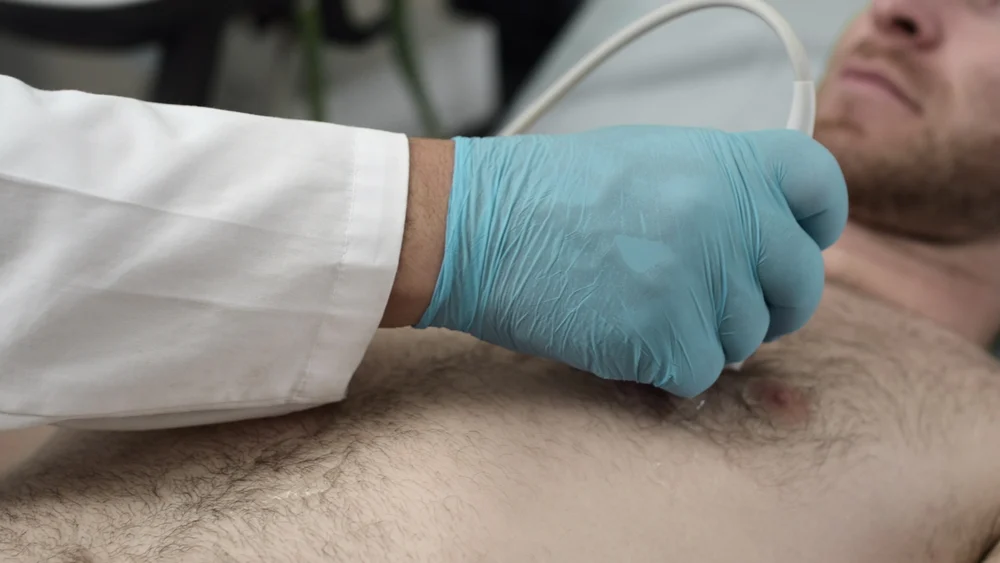
A study published in the Journal of Perinatology by researchers at the University of Chicago explored whether lung ultrasound (LUS) scores could serve as a reliable prognostic indicator. The investigation monitored 45 VLBW infants undergoing 53 extubation attempts following treatment for respiratory distress syndrome.
The methodology involved performing an LUS examination three to six hours prior to every extubation attempt. The researchers utilized a neonatal-adapted LUS scoring system to grade the state of the infants’ lungs. A failed extubation was strictly defined as any instance requiring reintubation within a seven-day window.
Key Findings and Clinical Utility
The data revealed that the neonatal-adapted LUS score is an exceptional predictor of success. By providing clinicians with a real-time visual assessment of lung aeration and fluid presence, the LUS score offers a higher degree of certainty than clinical observation alone. Furthermore, the researchers noted that common therapeutic interventions, such as dexamethasone, did not artificially inflate or deflate these scores, confirming the robustness of the LUS metric across varying treatment plans. This predictive capability allows neonatal intensive care units (NICUs) to personalize weaning protocols, minimizing the duration of invasive mechanical ventilation for the most vulnerable patients.
3. Combating Inappropriate Antibiotics with AI-Driven Diagnostics
The Antibiotic Stewardship Crisis
Lower respiratory tract infections (LRTIs) are among the most common reasons for antibiotic prescription in critically ill patients. However, distinguishing between bacterial infections and non-infectious causes of lung inflammation is notoriously difficult. This uncertainty often leads to the over-prescription of antibiotics, fueling the global crisis of antimicrobial resistance.
A Multimodal Diagnostic Approach
A collaborative team at the University of California, San Francisco (UCSF), has developed an innovative diagnostic model that integrates host biology with generative AI. The study, published in Nature Communications, centers on the identification of FABP4—a gene that, when expressed in lung fluid, serves as a natural anti-inflammatory biomarker. In healthy lung cells, FABP4 levels are elevated, but in the presence of an LRTI, expression levels plummet.
The UCSF team combined this biological data with a generative AI analysis of electronic medical records (EMR), specifically training the model to synthesize information from chest X-ray reports and physician clinical notes. This "multimodal" approach mimics the way an expert clinician synthesizes various streams of information to form a diagnosis.
Performance and Future Directions
In an observational study of critically ill adults, the AI model achieved a 96% accuracy rate in diagnosing LRTIs, consistently outperforming experienced ICU clinicians. The most striking finding was the model’s potential to reduce inappropriate antibiotic use by more than 80%.
The authors emphasize that the model is designed for accessibility; it can be deployed via a HIPAA-compliant GPT-4 interface, allowing clinicians to receive rapid, data-backed support at the bedside. The UCSF team is now moving toward clinical validation and is exploring whether this methodology can be adapted to diagnose sepsis, a condition where the speed of accurate diagnosis is the primary determinant of patient survival.
Synthesis of Implications: The Future of Diagnostic Medicine
The Chronology of Innovation
These three studies—published in quick succession in early 2026—represent a shift from "reactive" to "proactive" medicine.
- 2025/2026: The focus has shifted toward integrating deep learning models into standard hospital equipment (ECGs, EMRs, and ultrasound machines).
- The Next Phase: Researchers are now moving from retrospective training cohorts to prospective clinical trials, ensuring that these AI-augmented diagnostic tools maintain their efficacy in diverse, real-world hospital environments.
Official Responses and Ethical Considerations
Medical bodies and health systems have responded with cautious optimism. While the diagnostic accuracy of these models is impressive, the "black box" nature of some AI systems remains a subject of scrutiny. Experts emphasize that these tools are intended to serve as "decision support" rather than "decision makers."
"The goal is not to replace the physician’s intuition," noted one lead researcher, "but to provide them with a high-fidelity ‘second opinion’ derived from patterns that are beyond the reach of human perception, such as subtle electrical shifts in an ECG or the microscopic expression of a gene."
The Road Ahead
The integration of these technologies into standard practice will require updates to hospital IT infrastructure, clinician training, and, perhaps most importantly, regulatory oversight. As hospitals adopt these AI-enhanced diagnostic workflows, they must also ensure that the data used to train these models is representative of diverse patient populations to avoid algorithmic bias.
Ultimately, the confluence of genomics, advanced imaging, and generative AI is providing clinicians with a clearer window into the physiological state of their patients. By enabling earlier detection of COPD, safer extubation in infants, and more judicious antibiotic use in the ICU, these innovations are not merely technological novelties; they are essential tools for reducing the global burden of disease and ensuring that the right patient receives the right treatment at the right time.
For further reading, please refer to the primary research papers:
- COPD Screening via ECG: eBioMedicine DOI: 10.1016/j.ebiom.2025.106066
- LUS and Extubation Success: Journal of Perinatology DOI: 10.1038/s41372-025-02525-5
- AI-Driven LRTI Diagnosis: Nature Communications DOI: 10.1038/s41467-025-66218-5