







Introduction: Moving Beyond the "Straight Tube" Paradigm
For decades, the study of human vascular health has been constrained by a fundamental limitation: the reliance on simplified, uniform models. In traditional laboratory settings, blood vessels were represented as idealized, straight tubes—geometric abstractions that bore little resemblance to the chaotic, branching, and dynamic reality of the human circulatory system. While these models provided foundational knowledge, they fundamentally failed to capture the high-stakes environment where vascular diseases, such as aneurysms and stenoses, typically originate.
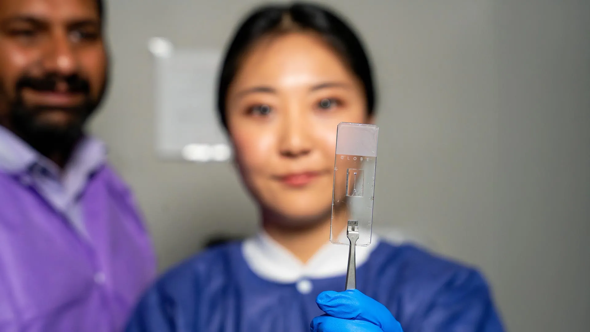
Researchers at the Texas A&M University Department of Biomedical Engineering have now shattered this paradigm. By developing a highly customizable, microfluidic "vessel-chip" system, the team in the Bioinspired Translational Microsystems Laboratory has bridged the gap between basic laboratory research and clinical reality. This advancement, recently published in the journal Lab on a Chip—and selected for the cover of the May 2025 issue—represents a significant leap forward in how we model disease, test pharmaceutical efficacy, and move toward a future of animal-free medical research.
The Chronology of Innovation: From Basic Concepts to Clinical Potential
The Genesis of the Vessel-Chip
The journey to this sophisticated technology began several years ago under the guidance of Dr. Abhishek Jain, an associate professor and the Barbara and Ralph Cox ’53 faculty fellow in biomedical engineering. The lab’s initial forays into the field focused on establishing the viability of microfluidic devices to mimic vascular behavior. Dr. Tanmay Mathur, a former graduate student, laid the essential groundwork by developing the first iteration of a "straight" vessel-chip. This early model proved that researchers could successfully cultivate living vascular cells within a controlled micro-environment, providing a proof-of-concept that would eventually lead to more complex architectures.
Jennifer Lee and the Evolution of Complexity
The project reached a new level of sophistication when Jennifer Lee, then an undergraduate honors student, joined the lab. Tasked with expanding upon the limitations of the straight-tube model, Lee sought to recreate the geometric irregularities that define the human body. Unlike a perfect cylinder, real-world vessels are characterized by bifurcations (branches), aneurysms (sudden expansions), and stenoses (narrowing).
"There are branched vessels, or aneurysms that have sudden expansion, and then stenosis that restricts the vessel," Lee explained. "All these different types of vessels cause the blood flow pattern to be significantly changed, and the inside of the blood vessel is affected by the level of shear stress caused by these flow patterns. That’s what we wanted to model."
Lee’s transition from a curious undergraduate to a lead researcher highlights the success of the lab’s mentorship model. By moving her project into the Master of Science fast-track program, the team was able to refine the chip’s design, optimize the flow mechanics, and eventually secure the high-level publication that validates the system for the broader scientific community.
Technical Foundations: The Science of Shear Stress and Architecture
At the heart of the vessel-chip is the concept of "fluid-structure interaction." In the human body, the physical shape of a vessel dictates how blood flows, and in turn, how the cells lining that vessel—the endothelial cells—respond to the physical force of that flow, known as shear stress.
When a vessel narrows (stenosis) or expands (aneurysm), the blood flow becomes turbulent or erratic. This altered physical environment is often the "trigger" for inflammatory processes that lead to disease. Previous models failed to account for this because they lacked the geometric complexity to generate these specific flow patterns.
The new Texas A&M vessel-chip utilizes advanced micro-fabrication techniques to create these precise shapes. By forcing fluid through these complex geometries, researchers can observe how endothelial cells react to localized stressors in real-time. This provides a "living" window into the pathogenesis of vascular diseases, allowing scientists to witness the onset of pathology in a way that was previously impossible without invasive human trials or imprecise animal models.
Official Responses and Expert Perspectives
The Vision of Dr. Abhishek Jain
Dr. Jain views this technology as a cornerstone for the "fourth dimension" of organ-on-a-chip research. "We can now start learning about vascular disease in ways we’ve never been able to before," he stated. "Not only can you make these structures complex, you can put actual cellular and tissue material inside them and make them living. These are the sites where vascular diseases tend to develop, so understanding them is critical."
Regarding the academic growth of his students, Jain emphasized that the lab’s culture is designed to push boundaries. "Jennifer demonstrated perseverance, curiosity, and creativity and started taking up research projects very quickly," Jain said. "Our fast-track program enables students like Jennifer to take on high-impact, high-risk research and not just do a science project, but take it all the way to its outcome and get it published."
The Student Perspective
For Jennifer Lee, the experience provided more than just a breakthrough in microfluidics. It provided a roadmap for professional development. "It’s such a good environment to interact with not only peers but also graduate students and postdoctoral researchers," Lee noted. "You’re able to learn teamwork and communication, work ethic, and just trying different things out. I think it’s such a valuable experience that students have available."
Implications: A New Era for Drug Discovery and Personalized Medicine
Revolutionizing Pharmaceutical Testing
The implications for the pharmaceutical industry are profound. Currently, drug development relies heavily on animal models, which are often poor predictors of human physiological responses, or clinical trials, which are expensive, time-consuming, and risky. Vessel-chips offer a "middle ground"—a platform that uses human-derived cells to test the safety and efficacy of new drugs in a human-relevant context. Because these chips can be tailored to individual patients, they also hold the potential to usher in an era of personalized medicine, where a treatment can be tested on a patient’s own "virtual" vascular system before it is administered in the clinic.
The "Fourth Dimension" of Research
The team is already looking toward the future. The current model features endothelial cells, but the next phase of development involves integrating multiple cell types—such as smooth muscle cells and immune cells—into the chip architecture. This would allow researchers to model the crosstalk between different tissues, simulating the complex biological environment of a functional blood vessel.
Dr. Jain characterizes this as the "fourth dimensionality" of organs-on-a-chip: moving beyond simple cellular presence to simulating the complex interaction of cells, flow, and architecture. This holistic approach is the new frontier in biomedical engineering, moving the field away from static observations and toward a dynamic, systems-biology approach.
Supporting Data and Institutional Collaboration
The success of this research is a testament to the power of interdisciplinary collaboration and sustained institutional support. The project was not built in a vacuum; it was supported by an impressive coalition of domestic and international funding bodies, including:
- The U.S. Army Medical Research Program
- NASA (which is keenly interested in how vascular systems behave in microgravity environments)
- The Biomedical Advanced Research and Development Authority (BARDA)
- The National Institutes of Health (NIH)
- The U.S. Food and Drug Administration (FDA)
- The National Science Foundation (NSF)
- Texas A&M University Office of Innovation Translational Investment Funds
This breadth of support reflects the strategic importance of vessel-on-a-chip technology. Whether for protecting astronauts in space, developing rapid responses to public health threats, or advancing general cardiovascular health, the technology developed in the Bioinspired Translational Microsystems Laboratory is positioned to become an essential tool in the 21st-century medical toolkit.
Conclusion: The Path Ahead
As Jennifer Lee moves forward in her career, she leaves behind a legacy of innovation that has fundamentally altered the trajectory of the lab’s work. The vessel-chip system is no longer just a theoretical experiment; it is a functioning, scalable, and highly accurate platform for scientific discovery.
The transition from the "straight tube" model to the complex, branching, and living architectures of the new vessel-chip signifies a broader shift in science: a move toward embracing complexity rather than ignoring it. By mimicking the chaotic beauty of the human circulatory system, Texas A&M researchers are not just observing vascular disease—they are finally beginning to understand it. As the technology continues to evolve, incorporating more complex cellular interactions and real-world geometries, it will undoubtedly become a cornerstone of future therapeutic breakthroughs, potentially saving countless lives by rendering the process of drug discovery safer, faster, and infinitely more human.