







In the late 19th century, a medical diagnosis captivated the attention of physicians across Europe and North America. It was a condition that explained a constellation of baffling symptoms—chronic abdominal pain, debilitating fatigue, and severe digestive distress. They called it "Glénard’s disease," named after the French physician Dr. Frantz Glénard, who in 1885 meticulously documented how the internal organs of his patients would, quite literally, sag from their anatomical moorings.
Yet, as the 20th century progressed, this diagnosis—clinically termed visceroptosis—all but vanished from the medical lexicon. Today, it exists in a state of clinical limbo, relegated to the periphery of mainstream medicine. However, for the growing population of patients diagnosed with Hypermobile Ehlers-Danlos syndrome (hEDS), this "forgotten" condition is staging a quiet, yet significant, comeback.
Main Facts: Defining the Displacement
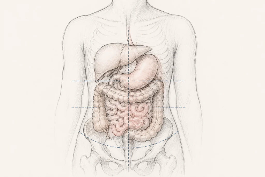
At its core, the term is etymologically straightforward. "Viscera" refers to the soft internal organs—the stomach, liver, intestines, and kidneys—housed within the abdominal and pelvic cavities. "Ptosis" denotes a downward displacement from the body’s normal anatomical alignment. Together, visceroptosis describes a state where gravity and a lack of structural support cause these organs to descend lower than they should.
While the human body is designed with a complex network of ligaments, mesenteries, and connective tissues to keep these organs suspended in their proper niches, this system is not infallible. In patients with connective tissue disorders, the very "glue" that holds the body together is structurally compromised. If the ligaments anchoring the stomach or the transverse colon become lax, the organs lose their fixed position, potentially leading to kinking, impaired blood flow, or obstructed transit.
A Chronological Journey: From Diagnosis to Dismissal
The history of visceroptosis serves as a fascinating case study in how medical trends dictate patient care. Between 1885 and the 1930s, the condition was a staple of prestigious journals such as The New England Journal of Medicine, JAMA, and The Lancet. Radiologists actively sought out "ptosis" during fluoroscopy, and surgeons developed specialized procedures to "tack" organs back into place.
Then, quite abruptly, the discourse stopped. The medical community began to view the condition with skepticism. Critics argued that organ position was highly variable and that "normal" was a subjective spectrum. Over the following decades, patients presenting with the classic symptoms of visceroptosis—bloating, early satiety, and chronic constipation—were increasingly categorized as having "nervous" or "psychosomatic" disorders.
The terminology shifted from physical diagnosis to moral judgment. Doctors began labeling these patients as "neurasthenic," "hysterical," or "anxious." This historical pivot meant that for over half a century, the structural reality of organ descent was ignored, and patients were frequently offered psychiatric referrals instead of diagnostic imaging. It is only in the last decade, through the lens of genetic connective tissue research, that the medical community has begun to revisit these early observations.
Supporting Data: The hEDS Connection
The modern resurgence of interest in visceroptosis is inextricably linked to the study of Hypermobile Ehlers-Danlos syndrome. In hEDS, the collagen that provides structural integrity to the body is defective. This systemic laxity explains why joint dislocations and subluxations are hallmark symptoms; it stands to reason that the internal "anchors" of the abdomen would be similarly affected.
The Spectrum of Symptomatic Descent
Modern clinical literature has begun to distinguish between anatomic visceroptosis (where an organ is lower than normal but functions perfectly) and clinical visceroptosis (where the descent causes functional impairment).
Research from the last few years highlights the gravity of this distinction:
- Gastrointestinal Impact: A 2023 study involving 139 patients with slow-transit constipation revealed a statistically significant correlation between transverse colon ptosis and the severity of bowel dysfunction. While this study was not exclusive to hEDS, it provided a robust clinical framework for understanding how physical displacement disrupts physiological function.
- The Pelvic Interface: Unlike abdominal visceroptosis, pelvic organ prolapse (POP) and rectal prolapse are well-documented in the 2017 diagnostic criteria for hEDS. This recognition acts as an "anchor" for further research; if the pelvic organs are demonstrably affected by collagen laxity, it is logically inconsistent to assume the abdominal organs above them remain immune to the same mechanical forces.
- The "Multifactorial" Hurdle: Despite this, medical consensus remains cautious. Gastrointestinal issues in hEDS are often labeled as "multifactorial," involving autonomic nervous system dysfunction, gut-brain axis disturbances, and motility issues. Disentangling whether a displaced stomach is the cause of nausea or merely a concomitant finding remains the primary challenge for clinicians.
Official Responses and Medical Perspectives
The medical establishment has been slow to formalize diagnostic protocols for visceroptosis. Part of this reluctance stems from a lack of standardized imaging. Standard GI evaluations—such as endoscopies or colonoscopies—are optimized to look for diseases inside the organs (like ulcers or tumors), not the position of the organs themselves.
Radiological studies designed to capture the organs in a functional, upright state are rare in modern practice. Consequently, many patients with symptomatic visceroptosis undergo extensive testing that returns "normal" results, reinforcing the frustrating cycle of medical gaslighting.
However, emerging voices in gastroenterology are calling for a shift. Proponents suggest that in patients with unexplained, refractory abdominal symptoms and known connective tissue laxity, "positional imaging" should be considered. By viewing the abdomen while the patient is upright, clinicians can finally visualize what was described in the 19th century: the physical collapse of the internal architecture.
Implications for Patients and Future Research
For the patient living with these symptoms, the implications of this knowledge gap are profound. The current reality is that patients must often act as their own medical detectives, pushing for imaging that is outside the standard of care.
The Path Forward: Treatment Strategies
Treatment remains largely conservative, focusing on managing symptoms while addressing the structural weakness:
- Physical Therapy: Specialized pelvic floor and core-stabilization physical therapy is the gold standard. By strengthening the musculature surrounding the abdominal cavity, patients may provide the external support their internal ligaments lack.
- Compression Therapy: The use of abdominal binders or medical-grade compression garments is frequently reported by the hEDS community as a means to alleviate the "heavy" or "dragging" sensation associated with organ descent.
- Dietary and Lifestyle Modification: Small, frequent meals to reduce the weight on the stomach and the use of specialized positioning during bowel movements can mitigate the impact of ptosis.
- Surgical Caution: Surgery remains a controversial topic. While some patients report success with "pexies" (surgical fixations), the systemic fragility of connective tissue in hEDS patients makes surgical intervention high-risk. Future research must prioritize identifying which patients are appropriate surgical candidates to avoid the complications of poor wound healing or recurrent prolapse.
Conclusion: A New Era of Understanding
The story of visceroptosis is a sobering reminder of the limitations of modern medicine. It demonstrates how easily a physical reality can be dismissed when it lacks a clear, high-tech diagnostic tool or a lucrative pharmaceutical solution.
As we move forward, the goal is not to return to the 1885 view of "Glénard’s disease," but to integrate historical clinical wisdom with modern structural insights. For the patient with hEDS, acknowledging the role of visceroptosis is not just about a label; it is about validating a lived experience. It is the transition from being told one’s pain is "all in the head" to recognizing that, for many, the source of the suffering is quite literally the weight of their own anatomy.
As research into connective tissue disorders matures, it is likely that visceroptosis will reclaim its place as a recognized, treatable clinical entity. Until then, the bridge between historical observation and modern, patient-centered care must continue to be built, one study—and one patient—at a time.
Key Takeaways
- Redefining History: Visceroptosis was a widely recognized condition in the late 1800s that fell out of favor, causing a gap in clinical knowledge that persists today.
- The hEDS Connection: Connective tissue laxity in hEDS provides a plausible biological mechanism for organ displacement that should be investigated further.
- Clinical vs. Anatomic: Not all displaced organs are symptomatic; diagnosis must focus on the correlation between displacement and functional impairment.
- The Need for New Standards: There is an urgent need for standardized imaging protocols that evaluate organ position in a functional, upright state.
- Conservative First: Physical therapy and abdominal support remain the safest initial interventions, with surgical options requiring extreme caution due to connective tissue fragility.