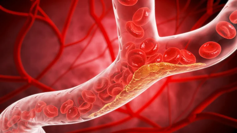


In the modern clinical landscape, chronic stress has long been regarded as a "silent epidemic"—an invisible force that quietly erodes cardiovascular health, weakens the immune system, and exacerbates conditions ranging from depression to obesity. Despite its pervasive nature, medical science has historically struggled to quantify it, relying on subjective patient questionnaires or cumbersome, point-in-time cortisol tests.
That is changing. Researchers at the Johns Hopkins University School of Medicine have developed a groundbreaking deep learning artificial intelligence model capable of identifying the first-ever biomarker of chronic stress visible on standard medical imaging. By analyzing the physical volume of the adrenal glands on routine chest CT scans, this AI tool provides a long-term, objective "gauge" of the body’s cumulative stress burden. The findings are slated to be presented next week at the annual meeting of the Radiological Society of North America (RSNA), marking a potential paradigm shift in how physicians identify patients at risk for stress-related cardiovascular disease.
The Science of Stress: Making the Invisible Visible
Chronic stress is not merely a state of mind; it is a physiological cascade. According to the American Psychological Association, the long-term activation of the body’s stress response system leads to sustained levels of cortisol and other hormones, contributing to high blood pressure, muscle pain, sleep disturbances, and a compromised immune system.
Until now, measuring this "allostatic load"—the cumulative wear and tear on the body—was a logistical challenge. Cortisol levels in the blood or saliva fluctuate wildly throughout the day, providing only a snapshot of a patient’s stress level at a single moment.
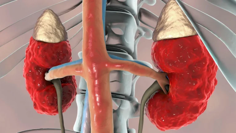
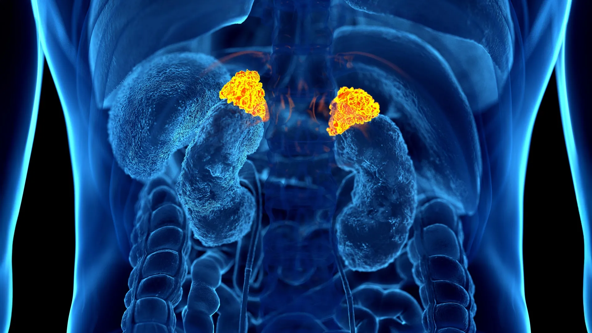
The research team, led by Dr. Elena Ghotbi, a postdoctoral research fellow at Johns Hopkins, recognized that the adrenal glands serve as the command center for the body’s stress response. As these glands work overtime to manage prolonged, chronic stress, they undergo physical changes. By training a deep learning model to automatically segment and measure the volume of these glands on standard chest CT scans—which are performed by the tens of millions annually in the United States—the researchers have created a way to look back in time at a patient’s physiological history.
Chronology of a Breakthrough
The path to this discovery involved an exhaustive integration of imaging, biochemistry, and psychosocial data. The project utilized the robust infrastructure of the Multi-Ethnic Study of Atherosclerosis (MESA), a massive, multi-decade research initiative.
- Data Synthesis (The Foundation): The researchers analyzed 2,842 participants (mean age 69.3, 51% women) who had undergone extensive baseline testing. This included chest CT imaging, validated psychological stress questionnaires, and rigorous salivary cortisol measurements taken eight times a day over two days.
- Developing the AI Model: Dr. Ghotbi and her colleagues trained a deep learning algorithm to identify and outline the adrenal glands on existing CT scans. They established the "Adrenal Volume Index" (AVI), a metric that accounts for body size by dividing adrenal volume (cm³) by height squared (m²).
- Cross-Validation: The team then compared the AVI scores against established "allostatic load" metrics, which included clinical markers like BMI, creatinine, hemoglobin, albumin, glucose, white blood cell counts, resting heart rate, and blood pressure.
- Longitudinal Correlation: Finally, the researchers mapped these findings against 10 years of follow-up clinical data to determine if higher AVI scores correlated with adverse cardiovascular events or mortality.
Supporting Data: The Link Between Volume and Vitality
The results were compelling, establishing a clear link between adrenal anatomy and systemic health. The AI-derived AVI values were found to be highly consistent with both psychological reporting and biochemical markers.
Participants who reported high levels of perceived stress—a subjective metric—demonstrated significantly higher AVI values compared to those who reported low stress. Furthermore, higher AVI values correlated with increased total cortisol exposure and a higher peak in cortisol levels throughout the day.
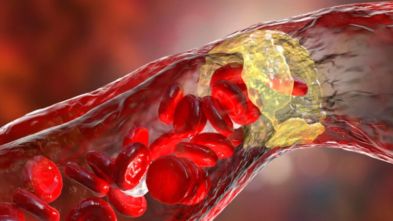
Perhaps most critically, the study established a clear clinical impact. Higher AVI was tied to a higher left ventricular mass index, an indicator of changes in heart structure that often precede heart failure. The data revealed a sobering trend: for every 1 cm³/m² increase in AVI, the risk of heart failure and subsequent mortality rose significantly. This makes the AVI not just an anatomical observation, but a validated, independent predictor of future clinical decline.
Official Responses and Expert Perspectives
The research has garnered significant attention from the medical community, with experts noting that this tool provides a long-sought bridge between radiology and preventative cardiology.
Dr. Shadpour Demehri, senior author and professor of radiology at Johns Hopkins, emphasized the practicality of the tool. "For the first time, we can ‘see’ the long-term burden of stress inside the body, using a scan that patients already get every day in hospitals across the country," Dr. Demehri stated. "Until now, we haven’t had a way to measure and quantify the cumulative effects of chronic stress, other than questionnaires or serum markers, which are very cumbersome to obtain."
Dr. Teresa E. Seeman, a professor of epidemiology at UCLA and a pioneering researcher in the field of stress and health, co-authored the study and highlighted its significance. "For over three decades, we’ve known that chronic stress can wear down the body across multiple systems," Dr. Seeman said. "What makes this work so exciting is that it links a routinely obtained imaging feature—adrenal volume—with validated biological and psychological measures of stress. It is a true step forward in operationalizing the cumulative impact of stress on health."
Dr. Ghotbi, the lead author, highlighted the efficiency of the model. "Our approach leverages widely available imaging data and opens the door to large-scale evaluations of the biological impact of chronic stress across a range of conditions," she said. "This AI-driven biomarker has the potential to enhance cardiovascular risk stratification and guide preventive care without additional testing or radiation."
Clinical Implications: The Future of Preventive Care
The primary implication of this discovery is the democratization of stress screening. Because the AVI can be calculated from CT scans already ordered for other reasons (such as lung cancer screenings or pulmonary evaluations), it requires no additional patient time, no extra radiation, and no additional cost to the healthcare system.
In a standard clinical setting, a radiologist’s report could eventually include an "Adrenal Volume Index," signaling to a primary care physician that a patient may be suffering from excessive, chronic stress. This allows for early intervention—such as lifestyle coaching, stress-reduction therapy, or more aggressive monitoring of blood pressure and cardiovascular health—long before a major heart event occurs.
Moreover, because the model is based on deep learning, its diagnostic accuracy is expected to improve as it is exposed to larger, more diverse datasets. While currently focused on middle-aged and older adults—a group at high risk for both cardiovascular disease and chronic stress-related complications—the researchers believe the model could eventually be adapted for a broader demographic.
As the RSNA meeting approaches, the medical community is bracing for what could be a foundational moment in diagnostic imaging. By turning a routine look at the body’s internal anatomy into a barometer for the mind’s cumulative burden, this AI model promises to turn the "invisible" epidemic of stress into a measurable, manageable, and treatable condition. In the eyes of the researchers, the goal is clear: to ensure that the "wear and tear" of life is no longer a silent killer, but a visible marker on the road to better, more personalized preventative care.