









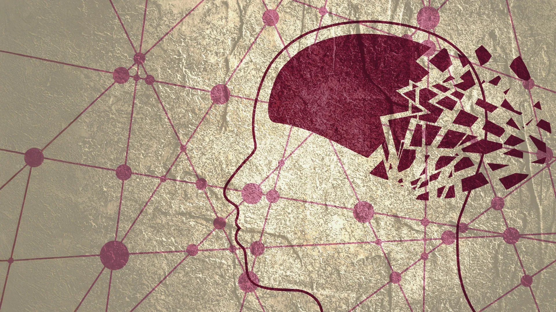
In the silent, intricate architecture of the human brain, a sophisticated plumbing system works tirelessly to flush out the toxic byproducts of daily neural activity. New research from Nanyang Technological University, Singapore (NTU Singapore) suggests that when these microscopic channels become obstructed, it may be one of the earliest "red flags" for the onset of Alzheimer’s disease.
For decades, the medical community has grappled with the challenge of diagnosing Alzheimer’s—the most prevalent form of dementia—only after significant cognitive decline has already taken root. Now, a groundbreaking study published by NTU’s Lee Kong Chian School of Medicine (LKCMedicine) indicates that "enlarged perivascular spaces" (EPVS)—the telltale signs of a clogged waste-removal system—can be detected via routine MRI scans long before the debilitating symptoms of memory loss and cognitive impairment become apparent.
The Anatomy of a Breakthrough: Understanding Perivascular Spaces
To understand the gravity of the NTU findings, one must first look at the brain’s waste management infrastructure. Perivascular spaces are fluid-filled channels that surround blood vessels. They serve as a critical drainage system, responsible for clearing out metabolic waste, including beta-amyloid and tau proteins—the very substances that aggregate to form the toxic plaques and tangles characteristic of Alzheimer’s.
When this drainage system is compromised, these channels expand, becoming visible as dark, fluid-filled pockets on standard magnetic resonance imaging (MRI) scans. While these enlargements have been noted in clinical settings before, their direct correlation with the underlying pathology of Alzheimer’s disease has remained largely speculative—until now.
By comparing these scans against clinical indicators and blood-based biochemical markers, the NTU research team has bridged the gap between structural brain changes and the molecular markers of dementia, suggesting that these "clogs" are not merely a byproduct of aging, but a proactive indicator of neurological decline.
Chronology of the Study: A Multi-Ethnic Perspective
The NTU research, conducted as part of the university’s rigorous Bachelor of Medicine and Bachelor of Surgery (MBBS) Scholarly Project module, was notable for its demographic scope. Led by Associate Professor Nagaendran Kandiah, the study analyzed nearly 1,000 participants across Singapore.
The research process unfolded over several critical phases:
- Cohort Selection: The team recruited a diverse group representing the multi-ethnic fabric of Singapore, including individuals with healthy cognitive function and those already experiencing mild cognitive impairment (MCI).
- Imaging Analysis: Participants underwent high-resolution MRI scans to map the density and distribution of perivascular spaces.
- Biochemical Correlation: Researchers collected blood samples to measure seven specific Alzheimer’s-related biochemicals, including amyloid-beta and tau proteins.
- Comparative Data Synthesis: The team analyzed the relationship between the physical obstructions (EPVS), established indicators of white matter damage, and the blood markers, establishing a clear hierarchy of predictive power.
The Vital Importance of Asian-Centric Research
One of the most compelling aspects of this study is its explicit focus on Asian populations. For too long, Alzheimer’s research has relied heavily on data from Caucasian cohorts. However, the biological drivers of dementia are not uniform across the globe.
"For example, among Caucasians with dementia, past studies show that the prevalence of a major risk gene, apolipoprotein E4, linked to Alzheimer’s is around 50 to 60 percent. But among Singapore dementia patients, it is less than 20 percent," explains Associate Professor Kandiah, who serves as the Director of the Dementia Research Centre (Singapore).
By diversifying the data set, the NTU team has demonstrated that the biomarkers and risk factors used in Western clinical practice may not always translate directly to Asian patients. This study underscores the urgent need for region-specific research to ensure that diagnostic tools are accurate and equitable for all populations.
Supporting Data: Connecting Clogs to Cognitive Decline
The study’s findings are bolstered by significant statistical correlations. The researchers observed that participants with mild cognitive impairment—the clinical "waiting room" for Alzheimer’s—were significantly more likely to exhibit enlarged perivascular spaces than their cognitively healthy counterparts.
Perhaps more striking was the comparison between these "clogged drains" and white matter damage. White matter damage (damage to the nerve fibers connecting brain regions) is a traditional marker used by clinicians to assess brain health. However, the NTU data revealed that, among individuals with MCI, the correlation between Alzheimer’s-related biochemicals and perivascular spaces was actually stronger than the connection to white matter damage.
This suggests that while white matter lesions are a useful indicator of general vascular health, enlarged perivascular spaces are a more specific, earlier harbinger of the molecular pathology unique to Alzheimer’s.
Official Responses and Clinical Perspectives
The medical community has received these findings with a mix of optimism and scientific rigor. Dr. Rachel Cheong Chin Yee, a Senior Consultant and Deputy Head at Khoo Teck Puat Hospital’s Department of Geriatric Medicine, emphasized the clinical utility of the discovery.
"These findings are significant because they suggest that brain scans showing enlarged perivascular spaces could potentially help identify people at higher risk of Alzheimer’s disease, even before symptoms appear," Dr. Cheong noted.
Similarly, Dr. Chong Yao Feng, a Consultant at the National University Hospital’s Division of Neurology, highlighted the shifting paradigm regarding cerebrovascular disease and Alzheimer’s. Traditionally, these have been treated as distinct clinical entities. The NTU study challenges this binary view. "The study’s findings are intriguing as they demonstrate that both diseases do interact in a synergistic manner," Dr. Chong explained.
He cautions, however, that clinicians must exercise balanced judgment. "Doctors will have to use their clinical judgement of the patient’s scan and symptoms, as well as discuss with the patient, to determine if more checks are needed to confirm whether a patient has Alzheimer’s disease or not."
Implications for Future Diagnosis and Intervention
The implications for the future of geriatric medicine are profound. If these enlarged spaces can be identified during routine health screenings—which many older adults already undergo—it provides a non-invasive, cost-effective "early warning system."
1. Early Intervention
Currently, by the time a patient presents with memory loss, a significant amount of neuronal damage has often already occurred. Identifying risk early allows for lifestyle interventions, medication management, and clinical trials that could potentially slow the progression of the disease.
2. Refining Clinical Protocols
The study encourages a shift in how radiologists and neurologists interpret MRIs. Rather than focusing solely on atrophy or white matter lesions, clinicians may soon be trained to monitor the health of perivascular spaces as a standard part of cognitive assessments.
3. A New Model of Disease Interaction
By viewing Alzheimer’s through the lens of vascular efficiency, the medical community may unlock new therapeutic targets. If the brain’s waste-removal system is "clogged," research into treatments that improve lymphatic drainage in the brain could become a new frontier in neurology.
The Path Forward: Longitudinal Verification
While the results are promising, the researchers are not resting on their laurels. The next chapter of this study involves longitudinal tracking. By monitoring the participants over the coming years, the team aims to determine the exact rate at which those with enlarged perivascular spaces progress to clinical dementia.
This follow-up phase is essential to validate the predictive power of these scans. If the data confirms that these obstructions are indeed a reliable indicator of future decline, the discovery could transition from an academic observation to a standard diagnostic tool in hospitals worldwide.
In the global fight against Alzheimer’s, where a cure remains elusive, the ability to "see" the disease coming has never been more vital. The work by NTU Singapore serves as a powerful reminder that sometimes, the most profound answers to complex diseases are hidden in the smallest, most overlooked corners of the human body. As the medical community continues to refine its approach to these "clogged drains," it offers a glimmer of hope for earlier detection, more informed clinical decisions, and a better quality of life for those at risk of dementia.