









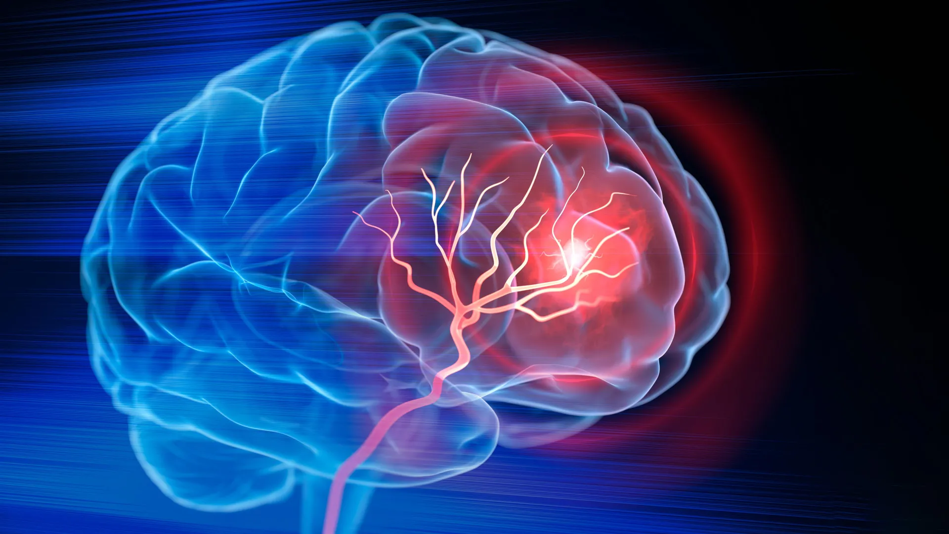
In the wake of an ischemic stroke, the brain is often left with a permanent, hollow scar—a physical void where healthy, functional tissue once thrived. For decades, the medical community has focused almost exclusively on the "golden hour" of acute intervention: the administration of clot-dissolving drugs and mechanical thrombectomies to restore blood flow. While these life-saving procedures preserve viable tissue, they cannot reverse the damage already done.
Now, a team of biomedical engineers at Duke University has unveiled a groundbreaking approach that shifts the focus from simple survival to active regeneration. By developing an injectable, porous biomaterial, researchers have successfully transformed the necrotic cavities left by strokes into fertile environments for neural and vascular repair. The study, recently published in the journal Cell Biomaterials, offers a glimmer of hope for restoring motor function long after the initial injury has occurred.
The Persistent Challenge of Post-Stroke Recovery
Ischemic strokes, which account for the vast majority of stroke cases, occur when a blood clot physically obstructs blood flow to a region of the brain. When neurons are deprived of oxygen and glucose for extended periods, they die. Once this tissue is lost, the resulting cavity presents a formidable obstacle to recovery.
"Once brain tissue has been lost, restoring blood flow is no longer enough," explains Tatiana Segura, the Robert Plonsey Distinguished Professor of Biomedical Engineering at Duke University. "Our goal is to engineer the injured space so that immune, vascular, and neural repair processes can begin to work together."
Currently, post-stroke recovery relies heavily on physical and occupational therapy. While these interventions help surviving brain circuits compensate for the lost tissue, they do not address the void itself. The brain, unlike the skin or liver, has a notoriously limited capacity for self-repair, often walling off damaged areas with scar tissue that physically and chemically prevents the growth of new connections.
The Engineering Strategy: MAPS and Molecular Signaling
To overcome these biological barriers, Dr. Segura’s team turned to a sophisticated material known as MAPS (Microporous Annealed Particle Scaffolds). MAPS consist of a collection of microscopic hydrogel particles that, when injected, self-assemble into a porous, three-dimensional structure. This structure acts as a temporary "scaffold," providing a physical architecture for cells to infiltrate and navigate.
However, the researchers realized that a structural framework alone was insufficient. To truly jump-start the repair process, they needed to communicate with the body’s own biological machinery. They focused their efforts on astrocytes—star-shaped glial cells that are the first responders to brain injury.
Harnessing Extracellular Vesicles (EVs)
Astrocytes communicate with their environment through extracellular vesicles (EVs)—tiny, nano-sized lipid packets filled with proteins, lipids, and genetic material. By harvesting these EVs from lab-grown astrocytes, the researchers could effectively "program" the scaffold.
Rather than diffusing these signals throughout the brain—where they might be diluted or trigger unintended side effects—the team chemically anchored the EVs directly onto the surfaces of the hydrogel microparticles. This concentrated the therapeutic signals within the stroke cavity, creating a localized, sustained "instruction manual" for any immune or neural cells that entered the scaffold.
Chronology of the Discovery: From Lab Bench to Functional Recovery
The research team’s approach was systematic, evolving from material design to functional validation in animal models.
- Material Optimization: The team first perfected the MAPS architecture, ensuring it was biocompatible and porous enough to allow cell infiltration.
- Signal Integration: They screened various signaling molecules to identify the most potent combination. They discovered that tethering Interleukin-4 (IL-4) and Complement Component 1q (C1q) to the scaffolds was particularly effective in recruiting beneficial immune cells.
- In Vivo Implementation: The optimized scaffold was injected into the stroke-affected regions of mice.
- Biological Response Monitoring: Over the course of eight weeks, the researchers monitored the recruitment of immune cells, the sprouting of new blood vessels, and the regeneration of axonal fibers.
- Behavioral Assessment: The final stage involved standardized grid-walking tests to measure the mice’s ability to coordinate their limbs, providing a direct correlation between the biological repair and behavioral improvement.
Supporting Data: The Unexpected Role of Neutrophils
One of the most provocative findings of the study concerns the role of neutrophils. Traditionally, neutrophils are viewed as the "villains" of the inflammatory response; their influx into the brain during the initial stages of a stroke is often associated with the expansion of damage and worsening of inflammation.
However, the Duke team’s findings suggest a nuanced shift in this narrative. When the researchers analyzed the environment inside the MAPS scaffold, they found a persistent population of neutrophils that appeared to be actively supporting tissue repair rather than inhibiting it.
"This result changes how we think about neutrophils after stroke," said Shangjing Xin, the study’s lead scientist and a postdoctoral fellow in the Segura Laboratory. "Their role appears to depend on when they arrive, where they are located, and the signals they receive from their surroundings."
To confirm this, the team performed a depletion study. When they reduced the neutrophil population, they observed a significant decline in blood vessel formation and a stalling of the scaffold’s remodeling. This confirmed that, in the context of the engineered environment, neutrophils were essential participants in the healing process.
Official Responses and Scientific Significance
The scientific community has noted the study’s focus on the microenvironment as a potential turning point for neuroregeneration. By focusing on the "ecology" of the injury rather than just the individual cells, the Duke team has moved toward a systems-biology approach to brain repair.
"We are not simply placing a material into the brain," Dr. Segura noted. "We are engineering a local environment that can coordinate several parts of the repair response."
The study also highlighted the vital importance of the scaffold itself. When the researchers attempted to inject the EVs without the supporting MAPS structure, the therapeutic effects were negligible. The porous architecture was essential to keep the signals concentrated and to provide the physical space required for vascular and neural remodeling. By eight weeks, the mice treated with the optimized scaffold showed such significant improvement in limb coordination that their performance was statistically indistinguishable from healthy control animals.
Implications for Future Human Therapies
While the results in murine models are promising, the researchers remain cautious about the path to human application. The current study utilized rat-derived astrocytes, but the team is already pivoting toward using human induced pluripotent stem cell (iPSC)-derived astrocytes. This transition is crucial for two reasons: it provides a scalable, sustainable source of EVs, and it allows for a higher degree of customization, potentially enabling doctors to tailor the EV signals to a patient’s specific injury profile.
The Path Forward
The journey toward clinical trials will involve several rigorous steps:
- Scaling and Safety: Testing the material in larger animal models to ensure that the inflammatory response and tissue remodeling remain safe and controlled.
- Delivery Logistics: Determining the optimal timing for intervention. While the current study suggests that the scaffold can repair chronic injuries, the window of opportunity for maximum efficacy remains a subject of active research.
- Regulatory Hurdles: Navigating the complex regulatory requirements for injectable, bioactive materials in the human central nervous system.
Dr. Segura remains optimistic about the philosophy behind the work. "You do not restore an ecosystem simply by containing the initial damage," she said. "You have to create the conditions that allow life to return. That is how we think about the stroke cavity. The material is not intended to reproduce the brain itself, but to create an environment where the body’s own cells can enter, communicate, and participate in rebuilding vascularized tissue."
By viewing the stroke cavity as an "injured ecosystem" rather than a dead zone, the Duke team has opened a new front in neuro-rehabilitation. If this research continues to show success in larger models, it could revolutionize how we treat the millions of individuals currently living with the long-term, debilitating effects of ischemic stroke. The dream of "rebuilding" the brain, once thought to be science fiction, is beginning to take shape within the tiny, porous confines of a hydrogel scaffold.