










For decades, the study of human vascular health was hampered by a fundamental disconnect between biology and engineering. While the human circulatory system is a masterpiece of fluid dynamics—characterized by intricate branches, sudden aneurysmal expansions, and restrictive stenoses—laboratory models were relegated to simple, straight, uniform tubes. These simplified conduits, while functional for basic testing, failed to capture the chaotic, high-pressure environments where the most critical vascular diseases take root.
Today, researchers at the Department of Biomedical Engineering at Texas A&M University are bridging that gap. By pioneering a customizable "vessel-chip" system, a team led by Dr. Abhishek Jain is transforming how we observe, analyze, and treat the pathologies of the human circulatory system. Their latest innovation, recently published in Lab on a Chip and slated for the cover of the journal’s May 2025 issue, represents a paradigm shift toward a more granular, patient-specific understanding of cardiovascular medicine.
The Problem with Traditional Modeling
To understand the significance of this breakthrough, one must first recognize the inherent limitations of standard laboratory practices. Blood vessels are not static pipes; they are dynamic, responsive tissues that react to the mechanical forces of blood flow, known as shear stress.
"There are branched vessels, or aneurysms that have sudden expansion, and then stenosis that restricts the vessel," explains Jennifer Lee, a master’s student in biomedical engineering and lead author of the study. "All these different types of vessels cause the blood flow pattern to be significantly changed, and the inside of the blood vessel is affected by the level of shear stress caused by these flow patterns. That’s what we wanted to model."
In straight, uniform tubes, shear stress is predictable and constant. In the human body, however, the geometry of a vessel dictates the local environment. A sudden narrowing—stenosis—creates turbulence that can trigger inflammation or plaque buildup. By ignoring these geometries, previous models effectively "smoothed over" the very conditions that researchers need to study to understand disease progression.
A Chronology of Innovation
The journey to the current vessel-chip system is a testament to the cumulative nature of scientific progress at Texas A&M’s Bioinspired Translational Microsystems Laboratory.
The foundation for this work was laid just a few years prior by Dr. Tanmay Mathur, a former graduate student under Dr. Abhishek Jain. Mathur successfully developed a "straight vessel-chip," a proof-of-concept device that demonstrated the feasibility of using microfluidics to replicate basic vascular functions. While a significant advancement, it remained a simplified model.
Jennifer Lee joined Dr. Jain’s lab as an undergraduate honors student, entering the field with little prior exposure to organs-on-a-chip technology. Recognizing her potential, Dr. Jain encouraged her to push the boundaries of Mathur’s original design. Through the university’s Master of Science fast-track program, Lee transitioned from an undergraduate learner to a lead researcher. She dedicated her efforts to refining the microfluidic architecture, moving away from the "straight pipe" paradigm toward a design capable of replicating the complex, irregular geometries of the human vascular tree.
The culmination of this research—a customizable, patient-specific, and anatomically accurate vessel-chip—represents a multi-year evolution of lab technology, moving from basic validation to high-fidelity simulation.
The Mechanics of the Vessel-Chip
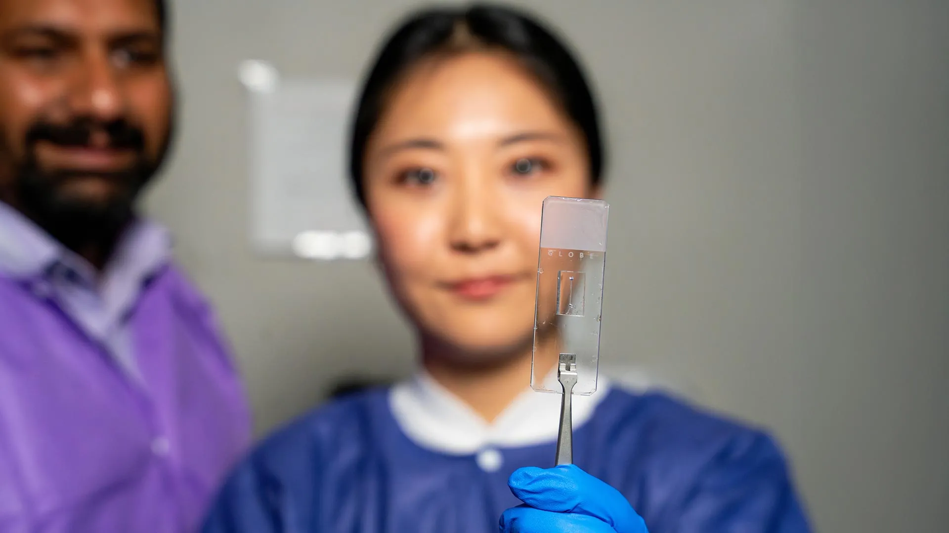
At its core, the vessel-chip is a microfluidic device, an engineering marvel that shrinks the scale of vascular anatomy to the microscopic level. These chips act as a non-animal alternative for medical research, providing a controlled, living environment where researchers can observe blood flow, introduce pharmaceuticals, and measure the biological response in real-time.
The "living" aspect of these chips is crucial. As Dr. Jain notes, "Not only can you make these structures complex, you can put actual cellular and tissue material inside them and make them living. These are the sites where vascular diseases tend to develop, so understanding them is critical."
By seeding the interior of these micro-channels with endothelial cells—the specialized cells that line the human blood vessel—the team creates a biological surrogate that mimics the responsiveness of a living human vessel. When blood (or a blood-like fluid) is introduced, the chip replicates the specific shear stresses and flow patterns found in the corresponding human anatomy, whether it is a healthy artery or a diseased vessel exhibiting narrowing.
Implications for Modern Medicine
The implications of this technology are vast, spanning from basic biological discovery to the rapid development of new therapeutics.
1. Precision Medicine and Patient-Specific Modeling
Because these chips can be tailored to the specific anatomy of individual patients, they open the door to "digital twin" or "physical twin" testing. Doctors could potentially extract cells from a patient, seed them into a chip modeled after that patient’s own vascular architecture, and test how specific drugs affect their unique disease presentation.
2. Drug Discovery and Toxicology
The current drug development pipeline is notoriously slow and expensive, often hindered by the high failure rate of drugs that perform well in simple petri dishes but fail in complex human systems. By providing a more accurate physiological model, the vessel-chip could allow pharmaceutical companies to filter out ineffective or toxic candidates much earlier in the process, reducing reliance on animal testing and accelerating the arrival of life-saving medicines.
3. The "Fourth Dimension" of Research
Dr. Jain describes the next phase of this research as the "fourth dimensionality of organs-on-a-chip." While traditional models have focused on cells (first dimension) and flow (second dimension), the inclusion of complex architectural states and the interaction of diverse cell types represents a new, multidimensional frontier. Future iterations of Lee’s design will incorporate multiple cell types beyond endothelial cells—such as smooth muscle cells or immune cells—to better capture the cross-talk between different tissues that defines vascular health.
Beyond the Lab Bench: Cultivating Future Scientists
While the technical output of the Bioinspired Translational Microsystems Laboratory is significant, the pedagogical impact is equally noteworthy. For Jennifer Lee, the experience served as a masterclass in professional development that transcended the technical requirements of the experiment.
"It’s such a good environment to interact with not only peers but also graduate students and postdoctoral researchers," Lee said. "You’re able to learn teamwork and communication, work ethic, and just trying different things out. I think it’s such a valuable experience that students have available."
Dr. Jain emphasizes that the success of the lab is predicated on the university’s fast-track program, which incentivizes students to pursue high-impact, high-risk projects. "Jennifer demonstrated perseverance, curiosity, and creativity," Jain said. "Our fast-track program enables students like Jennifer to take on sort of high-impact, high-risk research and not just do a science project, but take it all the way to its outcome and get it published."
A Multi-Institutional Effort
The development of the vessel-chip is not the result of isolated effort but a collaborative endeavor supported by a broad coalition of national health, military, and regulatory bodies. The project received critical backing from the U.S. Army Medical Research Program, NASA, the Biomedical Advanced Research and Development Authority (BARDA), the National Institutes of Health (NIH), the U.S. Food and Drug Administration (FDA), the National Science Foundation (NSF), and the Texas A&M University Office of Innovation Translational Investment Funds.
This wide-reaching support underscores the national importance of the technology. From NASA’s interest in how spaceflight affects human vasculature to the FDA’s interest in regulatory-grade alternatives to animal testing, the vessel-chip sits at the center of a major pivot in how the United States approaches medical research.
Conclusion: The Path Ahead
As the May 2025 issue of Lab on a Chip hits the presses, the work of Jennifer Lee and Dr. Abhishek Jain serves as a beacon for the future of biomedical engineering. By moving beyond the straight-line limitations of the past, they have unlocked a deeper, more nuanced way to view the human body.
The transition from a two-dimensional, uniform tube to a complex, living, and customizable vascular architecture is not merely a technical improvement; it is a fundamental shift in how we perceive the mechanisms of disease. As the team moves toward incorporating more cell types and refining the "fourth dimension" of their chip, the medical community waits with anticipation to see how these tiny, microfluidic devices will reshape the future of cardiovascular care, drug safety, and our fundamental understanding of the pathways of life.