







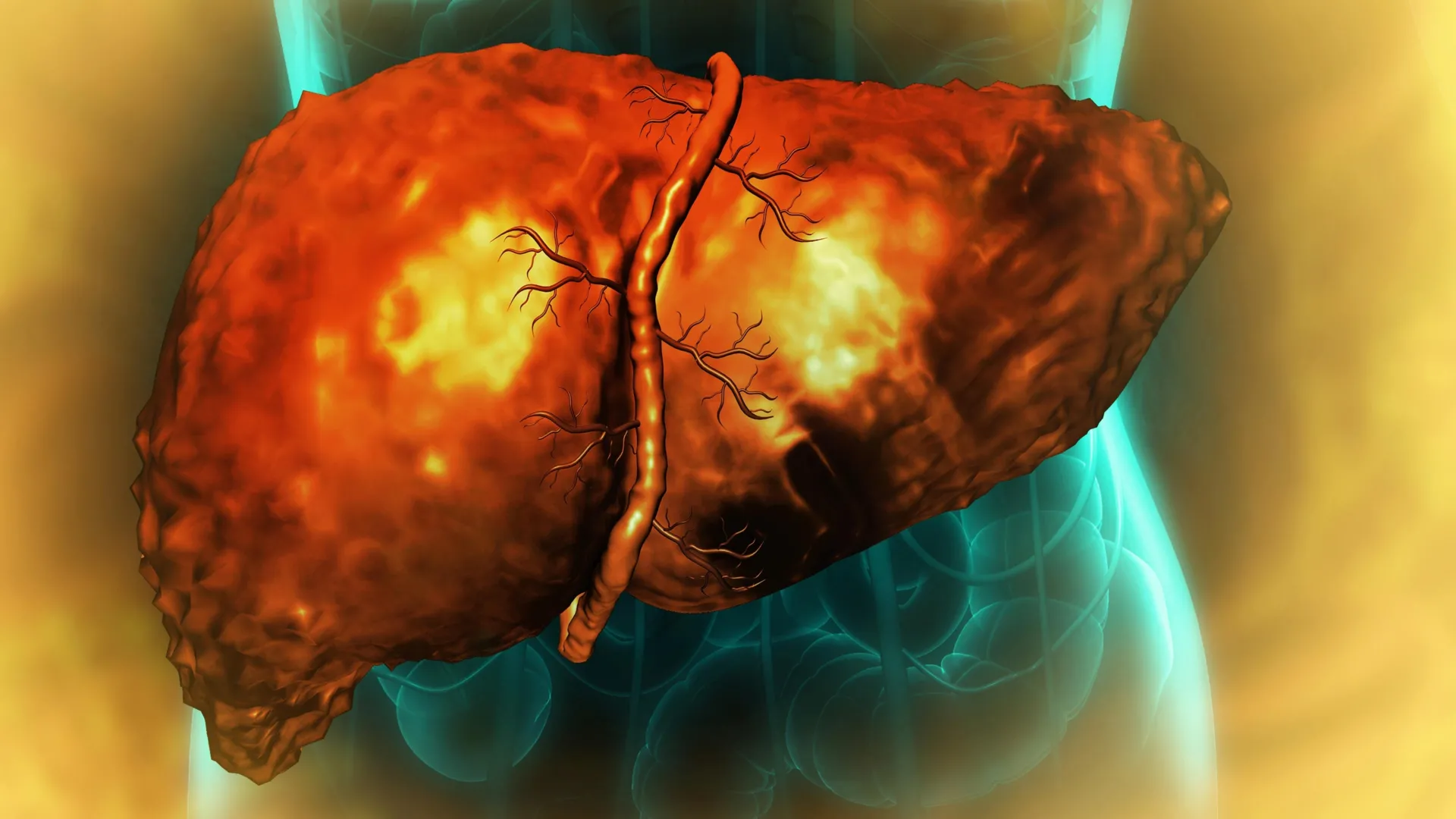
For most of human history, the liver has been regarded as a biological marvel—the only internal organ capable of near-complete regeneration. Even when severely damaged or partially excised, the liver possesses a sophisticated internal blueprint that allows it to revert, multiply, and mature back into a fully functional organ. However, for millions of individuals suffering from alcohol-associated liver disease (AALD), this evolutionary failsafe breaks down.
New, groundbreaking research published in the journal Nature Communications has finally identified the molecular "clog" that prevents this self-repair process. According to a collaborative study led by researchers at the University of Illinois Urbana-Champaign, Duke University, and the Chan Zuckerberg Biohub Chicago, alcohol-related damage leaves liver cells trapped in a pathological "middle state"—a cellular limbo where they are neither functional nor proliferative. This discovery offers a new understanding of liver failure and identifies specific biological pathways that could one day be targeted to reverse the damage, potentially reducing the need for life-saving, yet scarce, organ transplants.
The Anatomy of Regeneration: A Complex Biological Cycle
To understand the severity of this new discovery, one must first understand the liver’s standard operating procedure. Under normal conditions, the liver is a model of efficiency. When injury occurs, mature liver cells—known as hepatocytes—undergo a remarkable transformation. They temporarily shed their specialized identity and revert to a "progenitor-like" state. These cells are essentially stem-cell-adjacent; they possess the ability to divide and multiply, creating new tissue to replace what was lost. Once the tissue architecture is restored, these cells complete their maturation process, turning back into fully functional hepatocytes.
In patients with alcohol-associated hepatitis or cirrhosis, this elegant cycle grinds to a halt. The disease, which serves as a leading cause of global liver-related mortality and contributes to approximately 3 million deaths annually, creates a physiological environment where the liver simply loses its ability to heal itself. For decades, clinicians have observed that even after patients cease alcohol consumption, their livers often remain incapable of recovery. Until now, the underlying mechanism for this "point of no return" remained a mystery.
Chronology of a Discovery: Mapping the Molecular Breakdown
The research, led by U. of I. biochemistry professor Auinash Kalsotra and Duke University School of Medicine professor Anna Mae Diehl, sought to bridge the gap between clinical observation and molecular reality. The study began by analyzing tissue samples from patients with alcohol-associated hepatitis and cirrhosis, sourced through an initiative supported by the National Institutes of Health (NIH).
The "Quasi-Progenitor" Trap
As the research team compared these diseased tissues against healthy controls, a striking pattern emerged. The hepatocytes in diseased livers had indeed begun the transition toward the regenerative state, but they were unable to complete it. Instead, they remained suspended in an intermediate phase.
"They are neither functional adult cells nor proliferative progenitor cells," explained U. of I. graduate students Ullas Chembazhi and Sushant Bangru, the study’s co-first authors. "Since they are not functioning, more pressure builds on the remaining cells. They try to regenerate, and they all end up in this unproductive quasi-progenitor state, which is the driver of liver failure."
This cycle creates a downward spiral. As more cells enter this unproductive limbo, the burden on the remaining healthy cells increases. These healthy cells, triggered by the body’s distress signals, attempt to initiate repair, only to fall into the same trap, further depleting the organ’s functional capacity.
RNA Splicing: The Genetic Bottleneck
The breakthrough in the study came when the team moved beyond standard protein analysis and looked at the "intermediaries" of cellular function: RNA.
Genetic information in the DNA is transcribed into RNA, which then acts as a template for building proteins. Before this RNA can be used, however, it must undergo "splicing"—a process where non-coding segments are removed and essential pieces are joined together. This is a critical editing step; a single gene can produce different proteins depending on how its RNA is spliced.
Using deep RNA sequencing and advanced computational analysis, the researchers discovered that in alcohol-damaged livers, this splicing process was failing across thousands of genes. This was not a minor glitch; it was a systemic collapse of the cell’s "quality control" department.
The Role of ESRP2
The team identified the primary culprit as a deficiency in a protein known as ESRP2. Under normal circumstances, ESRP2 binds to RNA to ensure it is spliced correctly. In the presence of chronic alcohol-induced inflammation, ESRP2 levels plummet.
The consequences of this deficiency are twofold. First, the mis-spliced RNA leads to the production of dysfunctional proteins. Second, and perhaps more critically, the mis-splicing alters the "zip codes" of these proteins. Proteins are designed to function in specific locations—some in the cytoplasm, some in the nucleus. The researchers found that because of the errors in RNA splicing, vital regenerative proteins were being produced but were failing to reach the nucleus where they were needed to regulate gene expression. Instead, they were getting "stuck" in the cytoplasm, rendering them entirely useless.
Validating the Findings: From Mice to Medicine
To confirm that ESRP2 deficiency was truly the cause—and not just a symptom—of the failure, the researchers utilized mouse models. By breeding mice that lacked the gene responsible for ESRP2, they observed the same patterns of stunted regeneration and liver injury seen in human patients with advanced AALD.
The researchers then traced the source of the ESRP2 deficiency back to chronic inflammation. They found that when the liver is bombarded by alcohol, immune cells and structural cells release a surge of inflammatory cytokines and growth factors. These signals act as a suppressive force, effectively silencing the production of ESRP2.
In a promising laboratory demonstration, the team used a molecule to block the receptors for these inflammatory signals in cultured liver cells. The result was immediate: ESRP2 levels recovered, and RNA splicing returned to normal.
Implications for Future Clinical Care
The findings from Kalsotra and Diehl’s team provide a new roadmap for treating a disease that has historically left clinicians with few options beyond transplantation.
Diagnostic Potential
The researchers suggest that the specific "mis-spliced" RNA molecules could serve as powerful diagnostic biomarkers. By testing for these specific genetic signatures, physicians might be able to identify patients at high risk for liver failure much earlier in the disease progression, or monitor how well a patient is responding to lifestyle interventions.
Therapeutic Targets
The most exciting implication is the potential for new drug therapies. If scientists can develop treatments that either block the inflammatory signals suppressing ESRP2 or provide a way to bypass the need for ESRP2, it might be possible to "unlock" the trapped liver cells and kickstart the natural regenerative process. This would represent a paradigm shift in hepatology, moving away from reactive surgery toward proactive, molecular-level repair.
"I’m hopeful these findings will become a launching pad for future clinical studies," said Professor Kalsotra. "If we can correct the splicing defects, then maybe we can improve recovery and restore damaged livers."
The research team, which included contributors from the Carl R. Woese Institute for Genomic Biology, Johns Hopkins, and Northwestern, acknowledges that while the path from laboratory to bedside is long, the identification of the ESRP2 pathway provides the first tangible target for reversing the cellular, and potentially fatal, consequences of alcohol-associated liver disease.
Acknowledgments and Support
This extensive study was made possible through the support of the National Institutes of Health (NIH), the Chan-Zuckerberg Biohub Chicago, the Duke Endowment, and the Muscular Dystrophy Association. The research received funding through numerous NIH grants, including R01-AA010154 and R01-HL126845, reflecting the project’s significance to public health and the urgent need to address the global burden of liver disease.