









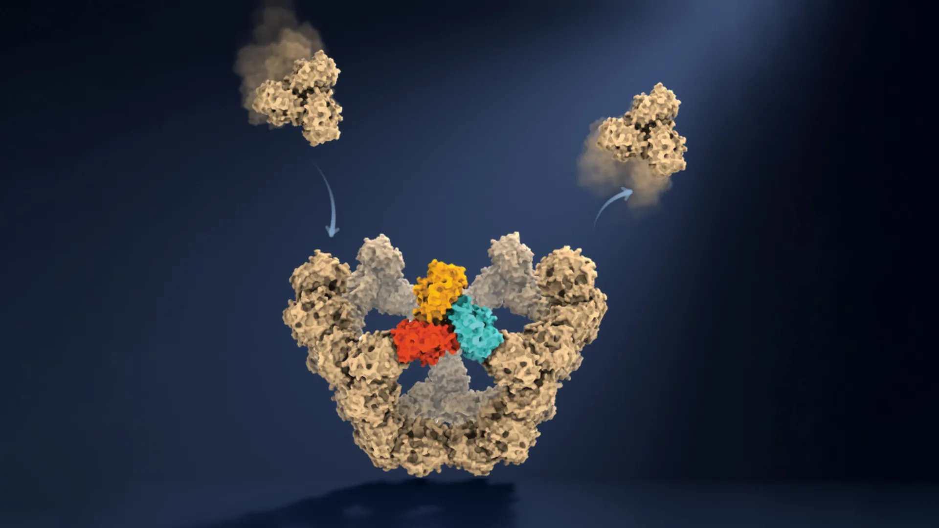
In a breakthrough that reads like a masterclass in molecular engineering, researchers at the University of Oxford have successfully observed the self-assembly of virus-like particles one molecule at a time. This milestone, published in the journal Nature, effectively demystifies the "hidden" rules governing how complex biological structures form with near-perfect reliability. For decades, the process of viral assembly—the mechanism by which a virus builds its protective protein shell, or capsid—remained a theoretical enigma. Now, by peering directly into the molecular forge, scientists have uncovered a surprisingly elegant system of trial, error, and structural "checkpoints" that guide chaotic interactions toward a precise, finished product.
The Architecture of the Invisible: Understanding Viral Assembly
Viruses are, in their simplest form, packages of genetic information encased in a protective protein shell known as a capsid. These shells are marvels of biological efficiency, often consisting of tens, hundreds, or even thousands of individual protein subunits. Despite the complexity of these structures, they spontaneously self-assemble without the aid of cellular machinery or external blueprints.
For years, the scientific community has been baffled by this process. How do these proteins, in a crowded, noisy cellular environment, consistently find the "correct" configuration when faced with thousands of potential, yet incorrect, connections? The Oxford team’s discovery suggests that the answer lies not in a rigid set of instructions, but in a sophisticated, self-correcting feedback loop. By watching this process unfold at the single-molecule level, researchers have finally transitioned from making educated guesses based on static snapshots to observing the dynamic "dance" of molecular construction.
Chronology of Discovery: From Theory to Observation
The path to this discovery was paved by a technological innovation known as mass photometry. Developed at the University of Oxford, this technique allows researchers to determine the mass of individual molecules by measuring the precise amount of light they scatter. While mass photometry had previously been used to analyze molecules, the team faced a significant hurdle: capturing the fleeting, intermediate structures that exist for only a fraction of a second.
Phase 1: Overcoming the Resolution Gap
Traditional methods, such as X-ray crystallography or cryo-electron microscopy, provide high-resolution images of "before" and "after" states—the isolated proteins and the fully formed capsid. However, they struggle to capture the transient states in between. To solve this, the researchers designed an experiment focusing on an engineered virus-like particle comprised of 60 protein units. They created a specialized environment that kept individual molecules confined while remaining continuously observable.
Phase 2: Real-Time Monitoring
By integrating their confinement method with high-speed mass photometry, the team successfully tracked the mass of a growing particle as it added individual building blocks. This was akin to filming a construction site where the bricks, mortar, and steel beams seem to assemble themselves into a skyscraper. For the first time, researchers could track the growth trajectory of a single particle, logging the precise moments when new proteins locked into place.
Phase 3: Identifying the Milestones
The resulting data revealed a clear, chronological sequence of assembly. The process is not a linear march toward success; it is a series of "molecular checkpoints." The most critical of these is the formation of a closed pentagonal ring. This specific structure serves as the first highly stable "anchor" in the assembly process. Once this pentagonal ring forms, the particle reaches a state of stability that allows subsequent additions to occur with increasing speed, effectively accelerating the completion of the capsid.
The Surprising Trick: How Molecular "Mistakes" Lead to Precision
Perhaps the most profound insight from the Oxford study is the role of imperfection. The researchers found that viral assembly functions much like navigating a complex maze. Many of the protein-to-protein connections formed in the early stages are weak and unstable.
This inherent "weakness" is not a flaw, but a critical feature. Because the initial connections are fragile, incorrect or sub-optimal combinations are prone to breaking apart. This creates a natural filter: if a group of proteins begins to assemble in an incorrect geometry, the weak bonds simply dissociate, giving the components another chance to find the right path.
As Dr. Roi Asor, co-lead author of the study from the Department of Chemistry and the Kavli Institute for Nanoscience Discovery, explains: "A virus has to solve an extraordinary construction problem. Its components somehow have to find the right arrangement among a huge number of possibilities, without a blueprint or machinery directing the process. We can now watch what happens molecule by molecule and see the physical rules that make it possible."
This process of "trial and error" continues until the proteins hit a specific, stable configuration. Once the structural "lock" is engaged, the process becomes significantly more robust, and the particle gains the stability required to grow toward completion.
Expert Perspectives and Official Findings
The study represents a collaborative effort across departments at the University of Oxford, highlighting the intersection of chemistry, physics, and molecular biology.
Professor Philipp Kukura, co-lead author and a pioneer in mass photometry, noted the paradigm shift this technology represents: "Until now, much of our understanding of how these structures assemble has had to be reconstructed from snapshots or theoretical models. Being able to both quantify the underlying interactions and follow one particle as it grows changes that. We can see the important intermediate structures appear and deduce a detailed model of the construction process."
The sentiment was echoed by PhD student and co-first author Dan Loewenthal, who simplified the magnitude of the achievement: "This method lets us study the assembly process directly; we just take a video!"
The ability to move from static data points to a continuous, observational video format allows scientists to map the "energy landscape" of viral assembly, identifying which states are favorable and which are dead-ends.
Implications: A New Frontier for Medicine and Nanotechnology
The implications of this discovery extend far beyond the study of viruses. By understanding the "rules of the game" for viral assembly, researchers are opening doors to a new generation of medical and engineering applications.
1. Disrupting Viral Replication
If scientists can identify the exact "checkpoints" where viruses form their shells, they can develop antiviral drugs specifically designed to sabotage these transitions. By introducing small molecules that either block the formation of the initial pentagonal ring or lock the protein shell into a "deformed" state, doctors could effectively stop a virus from ever becoming infectious.
2. Vaccine Development
The ability to observe and control the self-assembly process is a boon for vaccine design. Many modern vaccines rely on virus-like particles (VLPs) to trigger an immune response. Understanding how to guide these proteins to assemble correctly in the lab will lead to higher-yield, more stable, and more effective vaccine production methods.
3. Material Science and Nanotechnology
Self-assembly is a fundamental principle of nature. It is the same process that governs how cells build their internal skeletons, how complex protein complexes function, and how biological compartments are organized. The ability to observe these processes at the single-molecule level provides a blueprint for synthetic biologists and nanotechnologists. By mimicking the "trial and error" rules observed in viral assembly, scientists may eventually be able to program synthetic materials to build themselves into specific, useful structures, leading to advances in everything from targeted drug delivery systems to new smart materials.
Conclusion
The work conducted at the University of Oxford represents a triumph of observational science. By combining the precision of mass photometry with the patience to track individual molecular interactions, the team has turned a mysterious "black box" into a transparent process.
As Dr. Jack Tan of the MRC Weatherall Institute of Molecular Medicine aptly put it: "The ability to understand these processes at the molecular level could have important applications in vaccine development and antivirals that disrupt viral assembly."
As researchers continue to refine these techniques, the "maze" of viral assembly will become increasingly well-mapped. What was once viewed as an impossible biological puzzle is now being seen for what it is: a masterclass in elegant, self-correcting engineering. This discovery not only sheds light on the origins of viral infection but also offers a glimpse into the foundational mechanisms that allow life to assemble, adapt, and evolve.