










Glioblastoma, the most aggressive and lethal form of adult brain cancer, has long frustrated oncologists. Its ability to infiltrate healthy brain tissue with terrifying efficiency makes surgical resection nearly impossible and recurrence almost inevitable. While researchers have spent decades mapping the genetic mutations and chemical signaling pathways that drive this malignancy, a critical factor has remained hidden in plain sight: the physical nature of the environment surrounding the tumor.
Recent research published in Microsystems & Nanoengineering has uncovered a startling reality—the fluid surrounding glioblastoma tumors is not a passive bystander. It is a dynamic, viscous environment that acts as a training ground, priming cancer cells to become more invasive, flexible, and resilient. By developing a breakthrough microfluidic platform, researchers from Chongqing General Hospital and Chongqing University have demonstrated that this thick fluid doesn’t just impede cancer cells; it actively remodels them, endowing them with a form of "mechanical memory" that persists long after the cells leave the tumor’s edge.
The Physics of Invasion: A New Frontier in Oncology
Traditionally, cancer research has focused on the "seed and soil" hypothesis, looking at the genetic mutations within the cell (the seed) and the stiffness of the extracellular matrix (the soil). However, the interstitial fluid—the liquid medium filling the spaces between cells—has been largely overlooked.
In the case of glioblastoma, the physical properties of this fluid are far from uniform. Observations indicate that the fluid at the tumor’s advancing periphery is approximately eight times more viscous than the fluid found in the necrotic, dead-cell-filled center. As cancer cells push outward from the tumor core into the healthy brain, they are essentially swimming upstream through a thick, high-resistance medium.
For years, scientists struggled to study this phenomenon in a lab setting. Conventional microfluidic chips—the standard for simulating cellular environments—are closed systems. These channels often restrict oxygen flow, introduce artificial wall-friction, and fail to sustain long-term cell culture. Because of these technical limitations, the scientific community had no way of knowing whether the cells were simply struggling against the fluid or if they were undergoing a fundamental biological transformation in response to it.
Chronology of a Scientific Breakthrough
The path to this discovery began with a shift in engineering philosophy. Realizing that existing closed-channel systems were insufficient, the team in Chongqing set out to create an "open" microfluidic platform.
The Development Phase
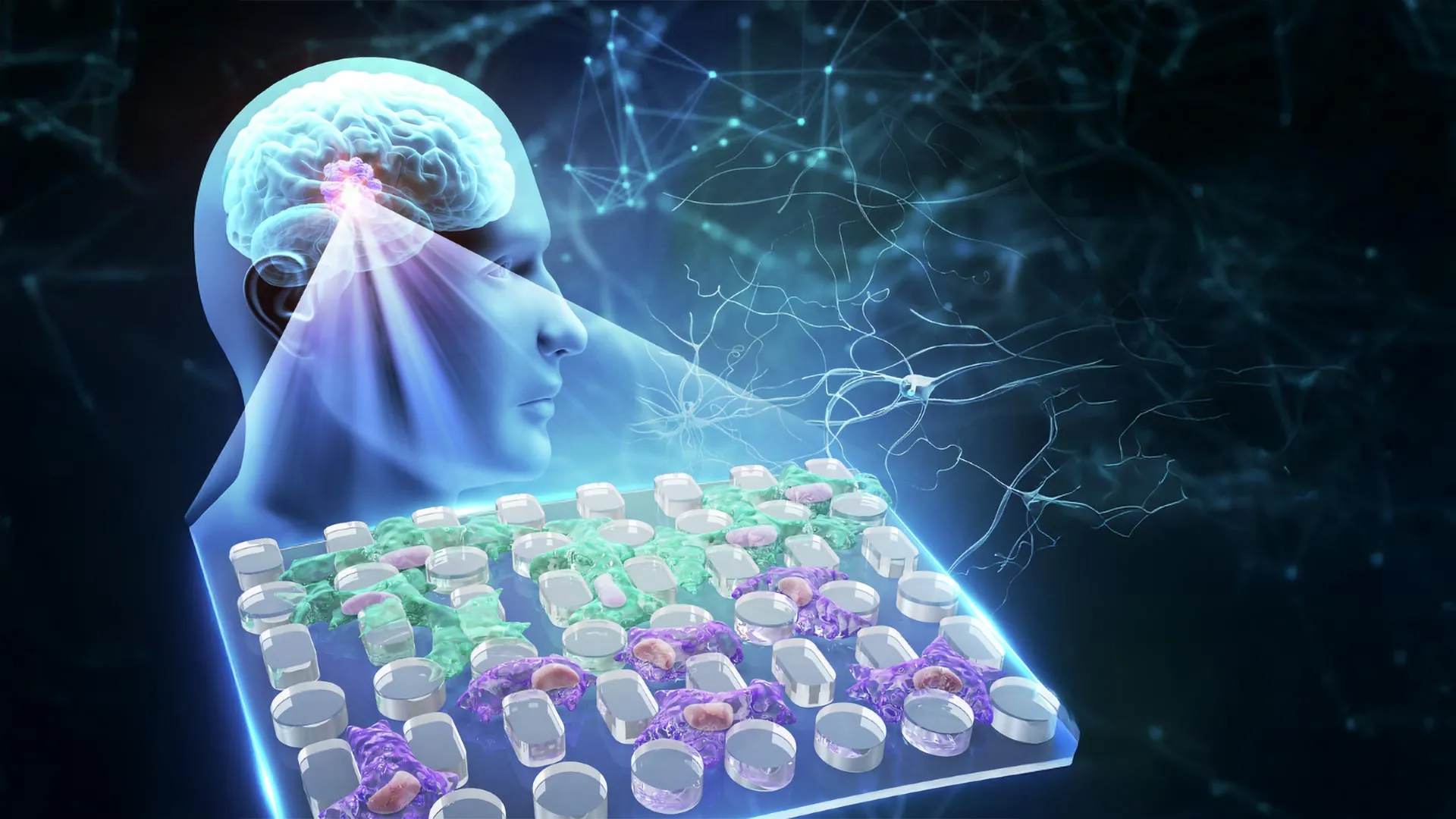
- Design Innovation: Researchers engineered a two-layer open microfluidic membrane equipped with a removable cap and a dense array of microscopic pillars. This design allowed for precise control over the cellular environment without the restrictive limitations of traditional closed conduits.
- Long-Term Cultivation: Unlike previous models that lasted only hours or days, the new device enabled the team to sustain human glioblastoma cell lines (U-251 and LN-229) in a controlled viscous environment for up to one month.
- Simulating the Tumor Edge: The team calibrated the fluid viscosity to 7.1 cP (centipoise), a measurement mirroring the conditions at the invasive front of a glioblastoma.
The Observation Phase
Once the cells were adapted to this viscous environment, the team subjected them to a series of stress tests. They observed the cells migrating through narrow gaps between the pillars to see how they would respond to physical confinement.
The results defied conventional intuition. Rather than being exhausted by the thick fluid, the "trained" cancer cells moved faster and covered more ground than their counterparts grown in standard, low-viscosity laboratory media. The fluid had not served as a barrier; it had served as a catalyst for evolution.
Supporting Data: Mechanical Adaptation and Molecular Shifts
The microscopic data provided a clear window into how these cells were changing. After weeks of exposure to high-viscosity fluid, the glioblastoma cells underwent distinct physical transformations:
- Size and Flexibility: The cells became smaller and significantly more deformable. This physical downsizing allowed them to squeeze through microscopic gaps that would have blocked ordinary cells.
- Nuclear Deformation: As the cells navigated the chip’s pillars, their nuclei—the most rigid part of a cell—showed increased plasticity, suggesting a restructuring of the internal cellular framework.
- YAP Signaling: Researchers detected an accumulation of YAP (Yes-associated protein) within the cell nuclei. YAP is a well-known mechanotransducer, a protein that acts as a sensor for physical stress. Its presence confirmed that the cells were not just reacting randomly, but were actively sensing the viscosity of their environment and triggering survival pathways.
Divergent Paths to Aggression
Perhaps the most intriguing aspect of the findings was the difference between the two cell lines. While both U-251 and LN-229 cells became more invasive, they took different biological routes to get there.
The U-251 cells underwent "mesenchymal-like reprogramming," a process where cells shed their stationary characteristics and adopt the migratory traits of mesenchymal stem cells. They specifically upregulated genes such as CD44, FN1, and MMP9—all of which are notorious for promoting cancer invasion and tissue remodeling.
In contrast, the LN-229 cells achieved similar levels of mobility without broad changes in gene expression. This suggests that glioblastoma cells possess a "redundancy" in their survival toolkit; if one path to invasion is unavailable, they can rely on structural flexibility rather than genetic modification to achieve the same lethal result.
The Concept of Cellular Memory
Perhaps the most significant revelation from the Chongqing team is the discovery of "mechanical memory." Through Western blotting, researchers confirmed that the protein changes induced by the viscous environment persisted even after the cells were transferred back into normal, low-viscosity fluid.
This implies that glioblastoma cells are not just responding to their immediate surroundings; they are recording their history. Once a cell has been "hardened" by the viscous fluid at the tumor edge, it carries that aggressive, highly mobile phenotype into the healthy brain tissue it invades. This permanent change means that the tumor’s "front line" is essentially a self-perpetuating force of cells that have been optimized for migration.
Implications for Clinical Oncology and Future Treatments
The discovery that physical environment drives cellular aggression opens a new chapter in cancer therapy. If viscosity is a key driver of metastasis, then our current approach to drug development—which relies heavily on chemical inhibition—may be missing a crucial target.
Rethinking Therapeutic Targets
The research suggests that drugs targeting YAP signaling or the cytoskeleton (the cell’s "skeleton") could be highly effective. By preventing the cell from "sensing" the viscous fluid or inhibiting its ability to become more flexible, clinicians might be able to lock the cancer cells in a less-mobile, more localized state.
A Tool for Precision Medicine
The new microfluidic platform is not just a research tool; it is a clinical candidate. Because the device is compatible with standard multi-well plates and allows for long-term imaging and staining, it could be used to screen patient-specific tumor samples. By testing how a patient’s unique glioblastoma cells respond to mechanical stress, doctors could potentially predict which tumors are most likely to be hyper-invasive, allowing for more aggressive or tailored interventions.
Limitations and Future Research
While these findings are groundbreaking, the researchers emphasize that this is a laboratory-based discovery. "We have identified the mechanism in a controlled environment," the research team noted. "The next step is to observe these behaviors in vivo and determine how this mechanical memory correlates with clinical outcomes in patients."
Conclusion: A Paradigm Shift
The study conducted at Chongqing General Hospital fundamentally challenges our understanding of brain cancer. It suggests that the "aggressiveness" of glioblastoma is not solely the result of genetic mutations or chemical signals; it is also a reaction to the physical architecture of the tumor’s microenvironment.
By recognizing that fluid viscosity is an active participant in tumor progression, scientists have uncovered a hidden vulnerability. If we can target the way cancer cells "learn" from their environment—and how they "remember" that training—we may finally be able to stop them from infiltrating the brain so effectively. In the complex battle against glioblastoma, the answer may not just lie in the code of the cell’s DNA, but in the physics of the fluid it calls home.
Funding and Support
This landmark study was made possible through the support of several key institutions. Funding was provided by the National Natural Science Foundation of China (grants 82473430, 22304143, 32501283), the Chongqing Talent Project (cstc2022ycjh-bgzxm0081), the Natural Science Foundation of Chongqing (CSTB2025NSCQ-GPX1077, CSTB2025NSCQ-GPX0309), the Young Medical Talent Program of Chongqing (YXQN2025088), the New Chongqing Project (CSTB2024YCJH-KYXM0115), the Research Funding of Chongqing General Hospital (2025022102), the Chongqing Special Funding for Postdoctoral Research Projects (2024CQBSHTB3131), and the Zhejiang Provincial Natural Science Foundation of China (LQN25E050020). These institutions, along with the Chongqing Research Center for Glioma Precision Medicine, continue to lead the way in uncovering the biological and physical mechanisms that drive this complex disease.