


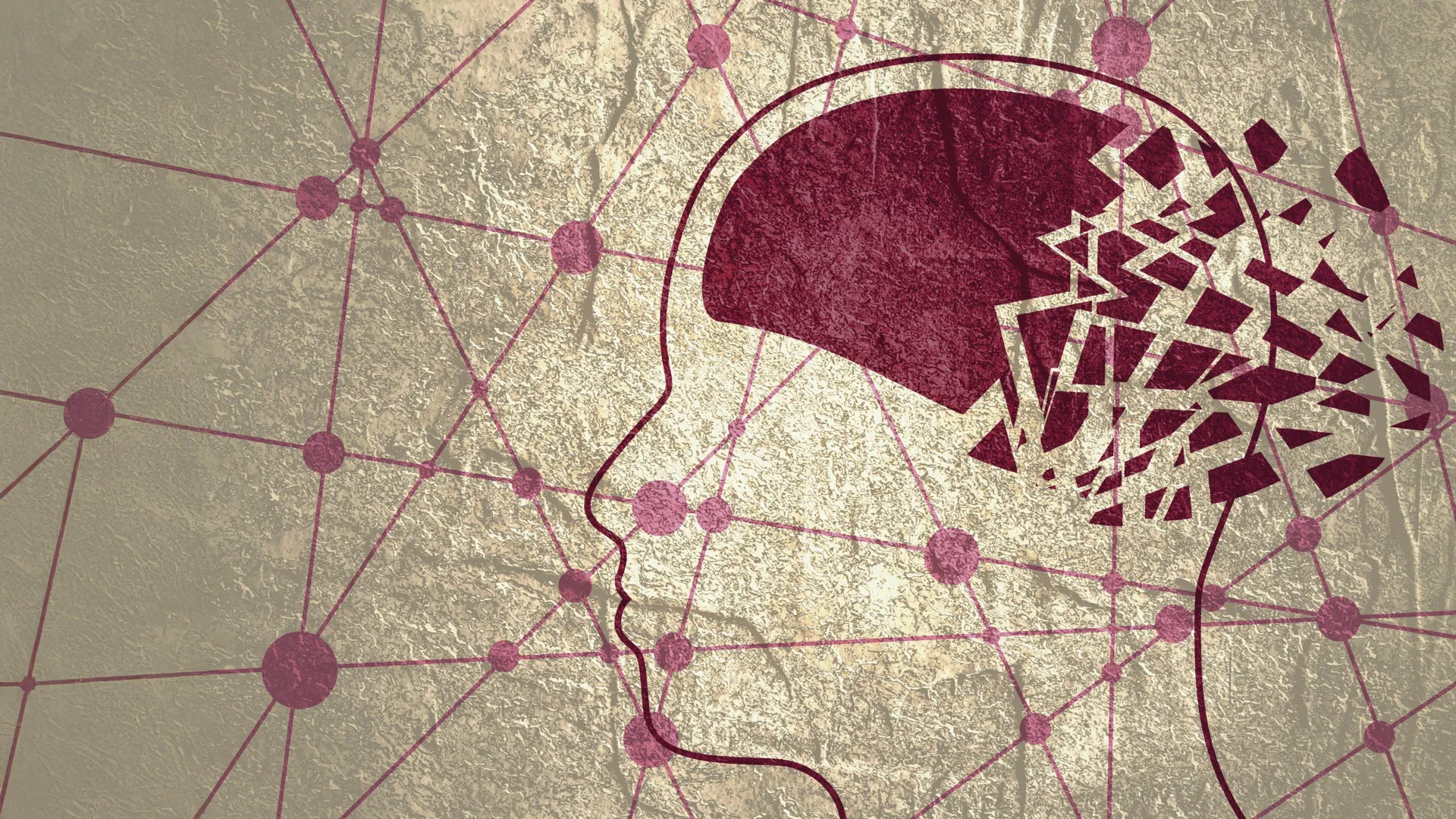
In the silent, complex architecture of the human brain, a sophisticated waste-removal system works tirelessly to clear out the biological "trash"—toxic proteins that, if left to accumulate, can trigger the devastating decline of Alzheimer’s disease. New research from Nanyang Technological University, Singapore (NTU Singapore), has uncovered a critical failure in this plumbing: when the brain’s waste-removal pathways become clogged, it serves as an early, visual warning sign of impending dementia, often manifesting long before cognitive symptoms become apparent.
This breakthrough, published by researchers at NTU’s Lee Kong Chian School of Medicine (LKCMedicine), suggests that a simple, non-invasive observation during a routine MRI scan could be the key to unlocking earlier interventions for one of the world’s most challenging medical conditions.
The Mechanics of Brain Health: Understanding Perivascular Spaces
To understand the significance of this discovery, one must first understand the brain’s unique anatomy. The brain does not have a conventional lymphatic system like the rest of the body. Instead, it relies on "perivascular spaces"—microscopic channels surrounding blood vessels—to flush out metabolic waste. These channels are responsible for clearing out harmful substances, most notably beta-amyloid and tau proteins.
In a healthy brain, these proteins are effectively washed away. However, as individuals age or develop neurodegenerative conditions, these pathways can become obstructed or enlarged. These "enlarged perivascular spaces" (ePVS) effectively act as a bottleneck, preventing the brain from cleaning itself. When these conduits become congested, the resulting buildup of toxic proteins can lead to the formation of amyloid plaques and tau tangles—the primary hallmarks of Alzheimer’s disease.
Bridging the Gap: A Study of Diverse Populations
A pivotal aspect of the NTU study is its demographic scope. Historically, the vast majority of Alzheimer’s research has focused on Caucasian populations in North America and Europe. This reliance on a single demographic has left a significant gap in our global understanding of the disease, particularly because Alzheimer’s manifests differently across ethnic and genetic backgrounds.
Associate Professor Nagaendran Kandiah, who led the research and serves as Director of the Dementia Research Centre (Singapore) at LKCMedicine, points to the stark contrast in genetic risk factors. "For example, among Caucasians with dementia, past studies show that the prevalence of a major risk gene, apolipoprotein E4, is around 50 to 60 percent. But among Singapore dementia patients, it is less than 20 percent," he explains.
By analyzing nearly 1,000 participants from Singapore’s multi-ethnic population, the research team ensured that their findings were robust and reflective of a broader reality. The cohort included a mix of individuals with normal cognitive function and those exhibiting signs of mild cognitive impairment (MCI), providing a comprehensive snapshot of the transition from healthy aging to the early stages of dementia.
Chronology of the Investigation
The study was conducted as part of LKCMedicine’s Scholarly Project module, an initiative within the Bachelor of Medicine and Bachelor of Surgery programme. The investigation followed a rigorous multi-stage methodology:
Phase 1: Imaging and Comparison
Researchers compared MRI scans of participants with healthy cognitive function against those with MCI. They looked specifically for the presence of enlarged perivascular spaces, assessing their correlation with established indicators of Alzheimer’s, such as damage to white matter—the "wiring" of the brain that facilitates communication between different regions.
Phase 2: Biochemical Correlation
To validate the MRI findings, the team measured seven specific Alzheimer’s-related biochemical markers in the participants’ blood, including levels of beta-amyloid and tau. This step was crucial; it moved the study from mere structural observation to a functional understanding of what was happening at a molecular level.
Phase 3: Longitudinal Analysis
The researchers analyzed the data to determine which markers—ePVS or white matter damage—were more strongly associated with the biochemical warning signs of Alzheimer’s. The results indicated that in patients with mild cognitive impairment, the link between biochemical markers and enlarged perivascular spaces was significantly stronger than the link with white matter damage.
Supporting Data: Why "Clogged Drains" Matter
The findings provide a compelling argument for shifting how we interpret neuroimaging. While white matter damage has long been the gold standard for evaluating potential dementia risk, the NTU study suggests it may not be the earliest indicator.
The study found that enlarged perivascular spaces were associated with four of the seven blood-based biochemical markers, suggesting that those with clogged "drains" are at a significantly higher risk of experiencing the cellular damage that characterizes Alzheimer’s.
"Although white matter damage is more widely used in clinical practice to evaluate for dementia, our results suggest that enlarged perivascular spaces may hold unique value in detecting early signs of Alzheimer’s disease," Assoc Prof Kandiah stated. This discovery effectively challenges the status quo, suggesting that radiologists and neurologists should pay closer attention to these specific vascular anomalies when reviewing scans of patients presenting with early memory concerns.
Official Responses and Clinical Perspectives
The medical community has received the findings with cautious optimism, acknowledging that this research could reshape diagnostic protocols.
Dr. Rachel Cheong Chin Yee, a Senior Consultant and Deputy Head at the Department of Geriatric Medicine at Khoo Teck Puat Hospital, emphasized the clinical utility of the discovery. "These findings are significant because they suggest that brain scans showing enlarged perivascular spaces could potentially help identify people at higher risk of Alzheimer’s disease, even before symptoms appear," she noted.
Dr. Chong Yao Feng, a Consultant at the National University Hospital’s Division of Neurology, highlighted the intersection of vascular and neurodegenerative medicine. Traditionally, doctors have treated cerebrovascular disease and Alzheimer’s as separate entities. "The study’s findings are intriguing as they demonstrate that both diseases do interact in a synergistic manner," Dr. Chong remarked. He warned, however, that the discovery requires nuanced clinical judgment. When doctors see these markers, they must not jump to conclusions; rather, they should view them as a signal to initiate a more comprehensive diagnostic conversation with the patient.
Implications for the Future of Alzheimer’s Care
The potential implications of this research are far-reaching, both for patients and healthcare systems globally.
1. Cost-Effective Early Detection
One of the most promising aspects of this study is its reliance on existing technology. Because enlarged perivascular spaces can be identified during routine MRI scans, there is no immediate need for expensive, specialized tests to identify this risk factor. This could democratize early detection, making it more accessible to patients in a variety of healthcare settings.
2. Time as a Resource
Justin Ong, the study’s first author and a fifth-year medical student, stressed the importance of time. "Identifying Alzheimer’s sooner gives doctors more time to intervene and potentially slow the progression of symptoms such as memory loss, reduced thinking speed, and mood changes," he said. By identifying patients while they are still in the "mild cognitive impairment" phase, clinicians have a greater window of opportunity to implement lifestyle changes, manage comorbidities, and provide appropriate care plans.
3. A Paradigm Shift in Treatment
The realization that vascular health and Alzheimer’s are deeply intertwined suggests that future treatments might need to target the brain’s waste-removal efficiency. If scientists can find ways to maintain the patency of perivascular spaces, they might be able to prevent the buildup of toxins before permanent brain damage occurs.
The Path Forward: What Comes Next?
While the NTU study provides a robust foundation, the researchers are not stopping here. The next phase of their work involves a longitudinal study, tracking the participants over several years to observe how many progress from mild cognitive impairment to full-blown Alzheimer’s dementia.
This follow-up is essential to confirm whether the presence of enlarged perivascular spaces is a predictive tool with high sensitivity and specificity. If the data continues to support the current findings, the inclusion of "ePVS assessment" in standard MRI reporting could become a global medical directive.
Furthermore, the team hopes that other researchers worldwide will replicate these findings in diverse populations. If consistent across different geographic and ethnic groups, the ability to "see" the brain’s plumbing issues will represent a massive leap forward in the global fight against dementia. By looking at the brain not just as a collection of neurons, but as a system of interconnected, physiological pathways, science is moving closer to a future where Alzheimer’s is no longer an inevitable decline, but a manageable, and perhaps even preventable, condition.