








For decades, the standard laboratory model for studying human blood vessels has been fundamentally flawed. Despite the biological reality that our vascular system is a chaotic, branching, and dynamic network of twists, turns, and varying diameters, scientific study has long relied on straight, uniform tubes to simulate these pathways. While these simplistic models provided a basic foundation for cardiovascular research, they failed to account for the physical conditions where the most dangerous vascular diseases—such as aneurysms and stenoses—actually take root.
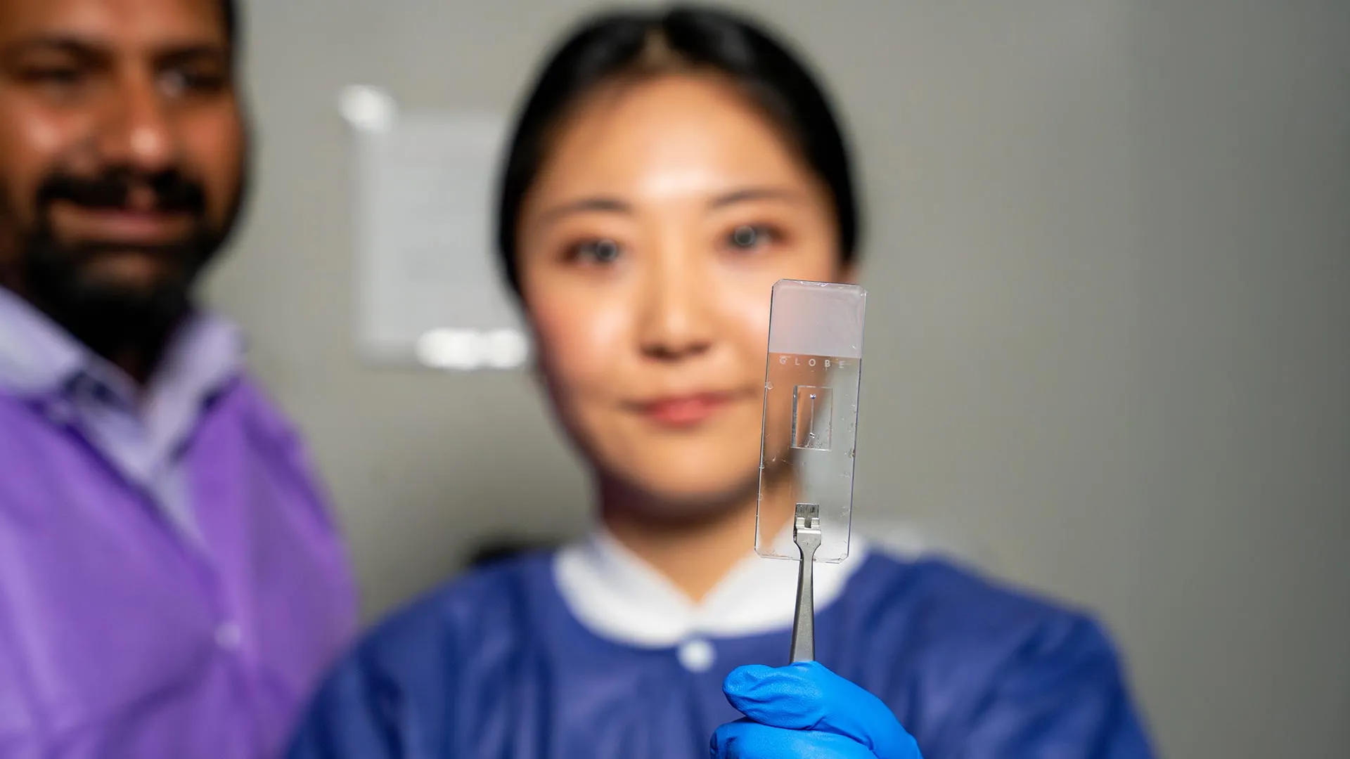
Now, a breakthrough from the Department of Biomedical Engineering at Texas A&M University is closing this gap. Researchers have unveiled a highly customizable "vessel-chip" system that moves beyond the limitations of legacy models, offering a realistic, microfluidic environment that mimics the complex architecture of human blood vessels. This advancement not only promises a more accurate understanding of how vascular diseases develop but also provides a powerful, non-animal platform for pharmaceutical testing.
The Science of Complexity: Redefining Vascular Architecture
The human vascular system is defined by its fluid dynamics. As blood flows through the body, it encounters complex geometries—bifurcations (branching points), sudden expansions like aneurysms, and constrictions known as stenoses. These structural irregularities are not merely incidental; they are the primary sites of shear stress—the frictional force exerted by blood flow against the vessel wall. It is precisely at these high-stress junctions where the lining of the blood vessel often begins to fail, leading to plaque buildup, clots, and chronic disease.
Jennifer Lee, a master’s student in biomedical engineering working in the Bioinspired Translational Microsystems Laboratory, led the design of the new vessel-chip. By utilizing advanced microfluidic fabrication, Lee created a system that can reproduce the wide variety of shapes found in human anatomy.
"There are branched vessels, or aneurysms that have sudden expansion, and then stenosis that restricts the vessel," Lee explained. "All these different types of vessels cause the blood flow pattern to be significantly changed, and the inside of the blood vessel is affected by the level of shear stress caused by these flow patterns. That’s what we wanted to model."
By replicating these specific flow patterns, researchers can observe how endothelial cells—the critical, thin layer of cells that line the interior of blood vessels—respond to physical stress in real-time. This provides a level of anatomical accuracy that was previously impossible to achieve in a laboratory setting.
A Chronology of Innovation: From Simple Tubes to Living Systems
The journey toward this breakthrough began several years ago in the lab of Dr. Abhishek Jain, an associate professor and the Barbara and Ralph Cox ’53 faculty fellow in biomedical engineering at Texas A&M.
The Foundation
The trajectory of this research was set in motion by Dr. Tanmay Mathur, a former graduate student in the Jain lab. Mathur developed the initial straight vessel-chip, a pioneering device that allowed researchers to observe blood flow under controlled, uniform conditions. While it served as a vital proof-of-concept, the team recognized that the "straight tube" limitation was a significant bottleneck in translational medicine.
The Developmental Phase
Jennifer Lee joined the lab as an undergraduate honors student with little prior experience in "organs-on-a-chip" technology. Under Dr. Jain’s mentorship, Lee began the rigorous process of refining microfluidic architectures. She transitioned from learning the basics of the technology to leading the design of a device capable of complex geometric manipulation. Her work successfully integrated these sophisticated shapes into the microfluidic platform, effectively moving the field from a one-dimensional view of vessel biology to a multidimensional one.
Recognition and Publication
The culmination of this research has garnered significant attention within the scientific community. Lee’s findings were published in the prestigious journal Lab on a Chip, and her work has been selected for the cover of the journal’s May 2025 issue, marking a significant milestone for a researcher who began the project as an undergraduate.
Official Perspectives: The Vision for Future Medicine
Dr. Abhishek Jain views the vessel-chip not just as a piece of hardware, but as a critical evolution in the field of regenerative medicine and drug discovery.
"We can now start learning about vascular disease in ways we’ve never been able to before," Dr. Jain said. "Not only can you make these structures complex, you can put actual cellular and tissue material inside them and make them living. These are the sites where vascular diseases tend to develop, so understanding them is critical."
The integration of living tissue into these chips is what distinguishes them from traditional synthetic models. By populating these chips with human endothelial cells, researchers create a "living" environment that can respond to pharmaceutical interventions. This allows for high-throughput testing of drugs on human cells, significantly reducing the reliance on animal models—a long-standing goal of the FDA and the broader scientific community.
Regarding the development of his students, Dr. Jain emphasized the value of the university’s fast-track program. "Jennifer demonstrated perseverance, curiosity, and creativity and started taking up research projects very quickly. Our fast-track program enables students like Jennifer to take on high-impact, high-risk research and not just do a science project, but take it all the way to its outcome and get it published."
Implications for Clinical Practice and Drug Development
The vessel-chip system carries profound implications for the future of personalized medicine. Because these devices are customizable, they can eventually be tailored to the unique biological profiles of individual patients. By using a patient’s own cells, clinicians could theoretically test how a specific individual’s vascular system reacts to different drug dosages, paving the way for truly personalized cardiovascular treatment.
Furthermore, the technology addresses the "fourth dimension" of organ-on-a-chip research. As Dr. Jain noted, the field is moving beyond just studying cells and flow in isolation; it is now entering an era where researchers examine the complex interplay between different tissue types and hemodynamic states.
"We are progressing and creating what we call the fourth dimensionality of organs-on-a-chip, where we not only focus on the cells and the flow, but this interaction of cells and flow in more complex architectural states, which is a new direction in the field," Jain added.
Future iterations of the vessel-chip are already in development. While the current model relies on endothelial cells, the research team is working to incorporate additional cell types, such as smooth muscle cells, to better replicate the multi-layered structure of human arteries and veins. This would allow for a deeper understanding of how different tissues communicate during the progression of diseases like atherosclerosis.
Beyond the Lab: Developing the Next Generation of Scientists
The impact of the Bioinspired Translational Microsystems Laboratory extends beyond the data collected in the chips. For students like Lee, the lab serves as a professional training ground. The environment emphasizes collaboration, technical communication, and the resilience required to navigate high-stakes scientific inquiry.
"It’s such a good environment to interact with not only peers but also graduate students and postdoctoral researchers," Lee said. "You’re able to learn teamwork and communication, work ethic, and just trying different things out. I think it’s such a valuable experience that students have available."
This interdisciplinary approach—blending rigorous engineering, cell biology, and professional development—is exactly what Texas A&M aims to foster in its engineering departments. By providing students with the opportunity to solve real-world problems that have tangible impacts on human health, the university is equipping them to lead the next generation of biomedical innovation.
Supporting the Future of Vascular Research
The significance of this research is underscored by the breadth of support it has received from both federal agencies and specialized research organizations. The project’s funding reflects its potential to revolutionize public health, with backing from the U.S. Army Medical Research Program, NASA, the Biomedical Advanced Research and Development Authority (BARDA), the National Institutes of Health (NIH), the U.S. Food and Drug Administration (FDA), the National Science Foundation (NSF), and the Texas A&M University Office of Innovation Translational Investment Funds.
This broad coalition of support highlights the versatility of the technology—from the FDA’s interest in reducing animal testing to NASA’s potential need for monitoring cardiovascular health in microgravity environments. As the technology matures, it is likely to become an indispensable tool in the fight against cardiovascular disease, the leading cause of death globally.
By challenging the "straight tube" paradigm and embracing the chaotic beauty of human biology, the team at Texas A&M has provided a roadmap for the future of vascular research. Through these tiny, complex, living chips, the medical community may soon gain the clarity needed to conquer some of the most persistent diseases of the human heart and circulatory system.