


For decades, chronic stress has been described as a "silent killer," a physiological specter that wreaks havoc on the human body while remaining notoriously difficult to quantify. Physicians have long relied on subjective questionnaires, cumbersome cortisol testing, or indirect markers of inflammation to gauge a patient’s stress load. Now, a team of researchers from the Johns Hopkins University School of Medicine has achieved a medical milestone: they have successfully utilized deep learning artificial intelligence to identify the first physical biomarker of chronic stress visible on standard medical imaging.
The breakthrough, which is scheduled to be presented at the upcoming annual meeting of the Radiological Society of North America (RSNA), promises to transform how healthcare providers assess the cumulative impact of stress on long-term patient health, turning routine chest CT scans into powerful diagnostic tools for psychological and physical well-being.
The Science of Visible Stress: Main Facts
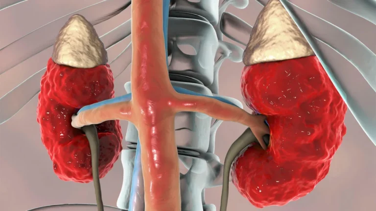
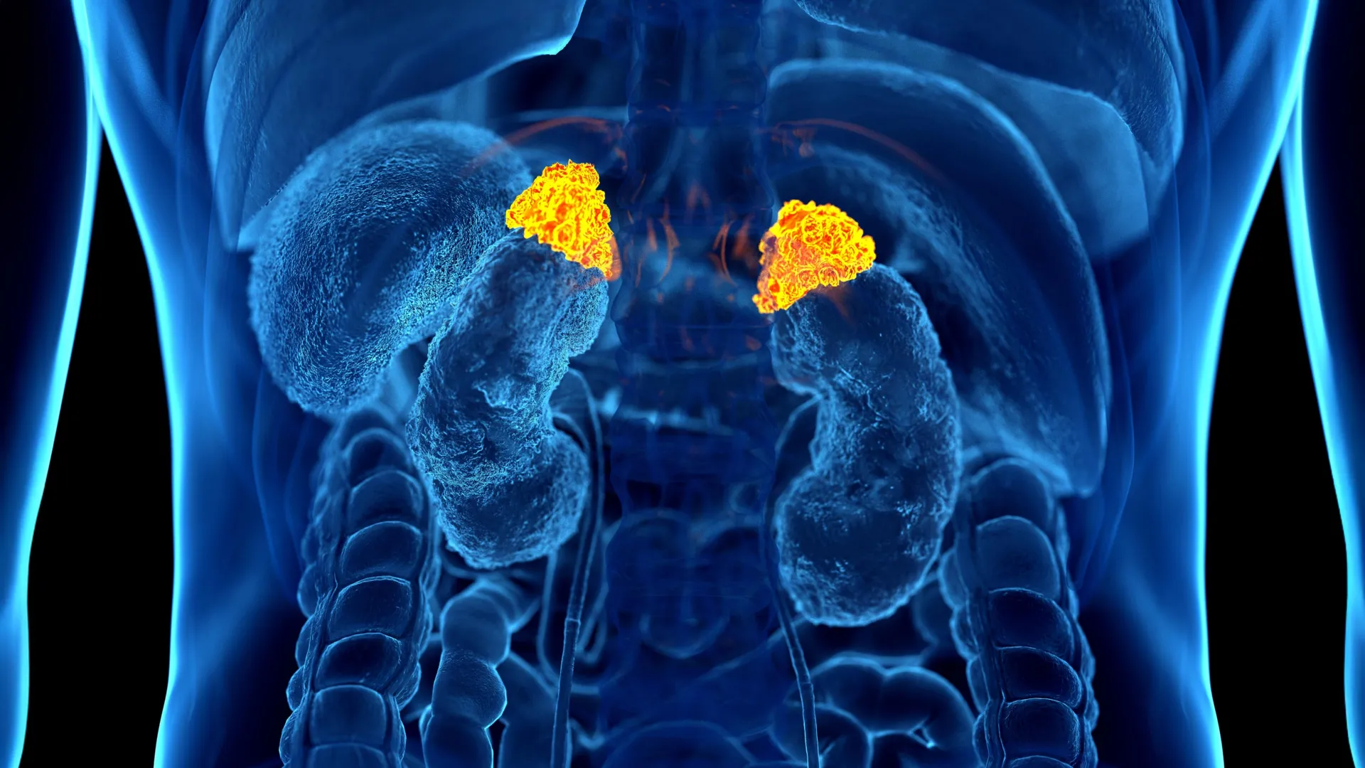
At the heart of this innovation is the Adrenal Volume Index (AVI). The adrenal glands, small, triangular-shaped organs located atop the kidneys, are the primary command centers for the body’s stress response. They release cortisol—the "stress hormone"—in response to ongoing tension. When a person experiences chronic, prolonged stress, these glands can physically enlarge due to persistent hormonal activation.
By developing a deep learning model capable of automatically segmenting and measuring the volume of these glands on CT scans, researchers have created a way to visualize the "wear and tear" of stress. The AVI is calculated by dividing the adrenal volume (in cubic centimeters) by the square of the patient’s height (in meters squared). This index provides a normalized, objective gauge that reflects long-term physiological stress rather than a fleeting moment of anxiety.
A Chronological Evolution of Stress Research
The journey to this discovery began with the realization that existing methods for measuring chronic stress were fundamentally flawed. Cortisol levels in blood or saliva can fluctuate wildly based on the time of day, diet, or recent activity, providing only a snapshot of a patient’s state.
- The Foundation: Researchers looked to the Multi-Ethnic Study of Atherosclerosis (MESA), a comprehensive, long-running study that has collected a vast wealth of data, including chest CT imaging, validated psychological stress questionnaires, salivary cortisol measurements, and "allostatic load" markers—a complex calculation of the body’s cumulative physiological strain, including blood pressure, glucose, and immune system markers.
- Model Training: Dr. Elena Ghotbi and her team at Johns Hopkins trained a deep learning AI to recognize and measure the adrenal glands within thousands of existing CT scans. The team analyzed data from 2,842 participants with a mean age of 69.3.
- Integration and Validation: Once the AI could consistently extract the AVI, the researchers cross-referenced these values with the biochemical and psychological data already present in the MESA cohort.
- Outcome Correlation: The team tracked participant outcomes over a decade, discovering a startling link: higher AVI values were consistently associated with higher cortisol levels, increased allostatic load, and, most critically, a higher incidence of heart failure and mortality.
Supporting Data: The Quantitative Link
The robustness of the study lies in the convergence of disparate data points. The researchers found that the AVI generated by their AI model did not just match theoretical expectations; it mirrored established clinical indicators of cardiovascular risk.
- Hormonal Alignment: Participants with higher AVI values demonstrated higher overall cortisol exposure and higher peak cortisol levels.
- Psychological Correlation: The AI-derived index correlated strongly with self-reported stress levels gathered through validated questionnaires. Patients who perceived themselves to be under higher stress consistently showed larger adrenal volumes.
- Clinical Impact: The data revealed a clear trend regarding heart health. An increased AVI was connected to a higher left ventricular mass index, a key measure of heart structure. The study established that for every 1 cm³/m² increase in the Adrenal Volume Index, there was a statistically significant increase in the risk of heart failure and death.
Official Responses and Expert Perspectives
The research has garnered significant attention within the medical community for its potential to move stress assessment out of the realm of abstract psychology and into the domain of precision medicine.
Dr. Elena Ghotbi, lead author of the study, emphasized the logistical brilliance of the approach: "Our approach leverages widely available imaging data and opens the door to large-scale evaluations of the biological impact of chronic stress across a range of conditions using existing chest CT scans. This AI-driven biomarker has the potential to enhance cardiovascular risk stratification and guide preventive care without additional testing or radiation."
Senior author Dr. Shadpour Demehri, a professor of radiology at Johns Hopkins, highlighted the historical struggle of quantifying the intangible. "For the first time, we can ‘see’ the long-term burden of stress inside the body, using a scan that patients already get every day in hospitals across the country," he noted. "Until now, we haven’t had a way to measure and quantify the cumulative effects of chronic stress, other than questionnaires, surrogate serum markers like chronic inflammation, and cortisol measurement, which is very cumbersome to obtain."
Dr. Teresa E. Seeman, a co-author and professor of epidemiology at UCLA, who is considered a pioneer in the study of stress and health, praised the study’s operational efficiency. "What makes this work so exciting is that it links a routinely obtained imaging feature, adrenal volume, with validated biological and psychological measures of stress and shows that it independently predicts a major clinical outcome. It’s a true step forward in operationalizing the cumulative impact of stress on health."
Clinical Implications: The Future of Preventive Care
The implications of this technology for everyday clinical practice are profound. In the United States alone, tens of millions of chest CT scans are performed annually for various medical indications, ranging from cancer screening to pulmonary investigations. By integrating an automated AI analysis of these scans, hospitals could, in theory, generate a "stress report" as a standard part of a patient’s imaging readout without requiring the patient to undergo a single additional test.
Redefining Risk Stratification
Cardiovascular disease remains the leading cause of death globally. Currently, risk stratification focuses heavily on cholesterol, blood pressure, and smoking history. The inclusion of an "Adrenal Volume Index" could provide cardiologists with a new, critical piece of data regarding how a patient’s lifestyle and environment are physically manifesting in their organs.
A Scalable Tool for Public Health
Because the technology is software-based and utilizes existing imaging hardware, it is highly scalable. It does not require specialized, high-cost equipment, making it a viable candidate for widespread implementation in both urban hospital systems and smaller community clinics.
Beyond Heart Disease
While the current study focuses heavily on cardiovascular outcomes, the researchers suggest the biomarker could eventually be applied to a wider range of stress-related conditions. Chronic stress is linked to depression, obesity, insomnia, and weakened immune function. By quantifying the body’s physiological "stress debt," clinicians may be able to intervene earlier, prescribing lifestyle changes or therapeutic interventions before a patient reaches a crisis point.
Conclusion
The intersection of artificial intelligence and radiology has provided a window into the human body that was previously opaque. By transforming the adrenal glands into a gauge for chronic stress, the Johns Hopkins team has bridged the gap between subjective experience and objective medicine. As this technology moves from the research stage to potential clinical application, it offers a promising path toward a more comprehensive, compassionate, and precise approach to treating the cumulative toll of modern life.