









Introduction: The Geometry of Health
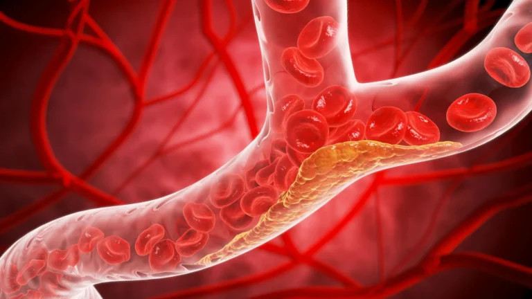
The human circulatory system is a masterpiece of biological engineering. Unlike the idealized, straight-walled conduits often depicted in medical textbooks, the reality of human vasculature is a labyrinth of complex geometries—branching arteries, serpentine veins, sudden aneurysmal expansions, and restrictive stenoses. For decades, however, the laboratory models used to study these pathways have been rudimentary, often treating blood vessels as simple, uniform cylinders.
While these simplified designs have provided a foundational baseline for cardiovascular research, they have historically failed to replicate the intricate fluid dynamics that trigger the onset of life-threatening diseases. Recognizing this critical disconnect between benchtop models and clinical reality, researchers at the Texas A&M University Department of Biomedical Engineering have pioneered a customizable "vessel-chip" system. This innovation marks a transformative shift in how scientists simulate, observe, and treat vascular pathologies.
The Core Innovation: Bridging the Gap in Vascular Modeling
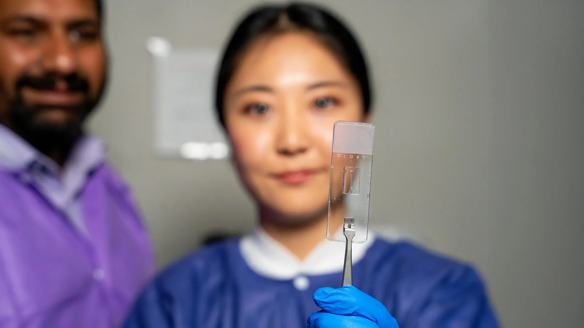
The vessel-chip, a hallmark of the burgeoning "organ-on-a-chip" field, is a microfluidic device engineered to replicate human blood vessels at a microscopic scale. By utilizing precision manufacturing, the team at the Bioinspired Translational Microsystems Laboratory has moved beyond the "straight-tube" paradigm to create structures that mimic the physical architecture of real-world anatomy.
The primary objective is to simulate the mechanical forces—specifically shear stress—that act upon the inner lining of vessels. Jennifer Lee, a master’s student in biomedical engineering who spearheaded the design, emphasizes that shape dictates function.
"There are branched vessels, or aneurysms that have sudden expansion, and then stenosis that restricts the vessel," Lee explains. "All these different types of vessels cause the blood flow pattern to be significantly changed, and the inside of the blood vessel is affected by the level of shear stress caused by these flow patterns. That’s what we wanted to model."
By replicating these complex shapes, the chips provide a high-fidelity environment where researchers can observe how cells react to irregular blood flow—a key factor in the development of atherosclerosis, thrombosis, and other vascular conditions.
A Chronology of Discovery: From Basic Models to Advanced Architecture
The path to this breakthrough was not immediate; it was a deliberate, iterative process fostered within the Bioinspired Translational Microsystems Laboratory, led by Dr. Abhishek Jain.
- The Foundational Phase: The project began with earlier, simpler iterations. A few years prior, Dr. Tanmay Mathur, then a graduate student in the lab, successfully developed a "straight vessel-chip" design. This initial proof-of-concept established that microfluidic environments could effectively sustain living vascular cells.
- The Expansion Phase: Building upon Mathur’s groundwork, Jennifer Lee joined the lab as an undergraduate honors student. Her tenure saw the integration of advanced manufacturing techniques capable of producing non-uniform vessel geometries.
- The Validation Phase: Through rigorous testing, the team proved that these complex chips could maintain cellular health while subjecting them to the variable fluid dynamics found in the human body.
- The Publication Milestone: The culmination of this research, which details the methodology for creating these complex architectures, was published in the prestigious journal Lab on a Chip. The study has earned the distinction of being featured on the cover of the journal’s May 2025 issue, signaling its significance to the international scientific community.
Official Perspectives: The Vision of Dr. Abhishek Jain
Dr. Abhishek Jain, an associate professor and the Barbara and Ralph Cox ’53 Faculty Fellow in Biomedical Engineering, views this development as a paradigm shift. According to Jain, the value lies not just in the plastic and silicon of the chip, but in its "living" potential.
"We can now start learning about vascular disease in ways we’ve never been able to before," Jain says. "Not only can you make these structures complex, you can put actual cellular and tissue material inside them and make them living. These are the sites where vascular diseases tend to develop, so understanding them is critical."
Jain describes the current work as part of an evolution toward the "fourth dimension" of organ-on-a-chip technology. While early iterations focused solely on cell viability or basic flow, the next generation of these devices will focus on the architectural context—the interaction of complex physical states with living tissue. This holistic approach is essential for drug discovery, as it allows pharmaceutical developers to see how a medication performs in a high-stress, anatomically accurate environment before it ever reaches a human trial.
Supporting Data and Technical Implications
The implications of this technology for the pharmaceutical industry are profound. Current drug development cycles are notoriously expensive and prone to failure, often because animal models—the traditional standard for preclinical testing—do not perfectly mirror human vascular physiology.
Key Advantages of the Vessel-Chip:
- Patient-Specific Customization: These chips can theoretically be tailored to reflect the unique anatomy of an individual patient, allowing for "personalized medicine" approaches where doctors can test which medication works best for a specific person’s vascular structure.
- Reduced Reliance on Animal Testing: By providing a more accurate human-centric model, these chips align with the growing global movement to reduce the use of animals in laboratory settings.
- Real-Time Visualization: Because the chips are transparent and micro-scale, researchers can use high-resolution imaging to watch the real-time response of endothelial cells—the cells that line the blood vessels—to various stimuli or drugs.
As it stands, the current model incorporates endothelial cells to form the vascular lining. However, the roadmap for the lab includes the introduction of secondary cell types, such as smooth muscle cells or immune cells, to recreate the complex tissue crosstalk found in the human cardiovascular system.
Professional Growth: The Human Element of Research
While the technical achievements are the headline, the project also serves as a case study for the value of academic mentorship. Jennifer Lee’s transition from an undergraduate newcomer to a published lead researcher is a testament to the infrastructure provided by the Texas A&M fast-track program.
"Jennifer demonstrated perseverance, curiosity, and creativity and started taking up research projects very quickly," says Dr. Jain. "Our fast-track program enables students like Jennifer to take on high-impact, high-risk research and not just do a science project, but take it all the way to its outcome and get it published."
For Lee, the experience was about more than just the data; it was about professional development. The collaborative environment of the lab, where she worked alongside post-doctoral fellows and senior researchers, allowed her to cultivate soft skills that are often overlooked in technical curricula—namely, team communication, project management, and the ability to pivot when experiments don’t go as planned.
"It’s such a good environment to interact with not only peers but also graduate students and postdoctoral researchers," Lee notes. "You’re able to learn teamwork and communication, work ethic, and just trying different things out. I think it’s such a valuable experience that students have available."
Conclusion: A New Horizon for Cardiovascular Medicine
The work being done in the Bioinspired Translational Microsystems Laboratory represents the confluence of mechanical engineering, cell biology, and clinical medicine. By moving beyond the simplistic "straight tube" models of the past, Texas A&M researchers are creating a new frontier in the study of vascular disease.
With robust backing from major funding bodies—including the U.S. Army Medical Research Program, NASA, the FDA, the National Institutes of Health, and the National Science Foundation—the project is positioned to significantly impact how we approach cardiovascular healthcare. As these living chips grow more complex, they promise to unlock a deeper understanding of human biology, ultimately accelerating the delivery of safer, more effective, and highly personalized medical treatments.
In the world of biomedical research, where every detail matters, the shift from simplicity to complexity is not just an academic upgrade—it is a necessity for saving lives.